Clinical Assessment
Traumatic optic neuropathy is a clinical diagnosis and should not be made if vision and pupillary function are normal. There must be a history of trauma. The clinical history should be as complete as possible. Commonly, vision will be 20/400 or less in the affected eye. The incidence of no light perception vision following TON varies significantly with most studies of 15 or more cases reporting an incidence that ranges from 22% to 78% ( Table 1 and Table 2 ).[12,42] However, visual acuity may only be slightly reduced. Recent series include many patients diagnosed with TON where vision in the affected eye was in the 20/25 to 20/70 range.[4,13,43] As vision tends to improve spontaneously following injury, the initial assessment should document the time interval between injury and the visual examination.
Delayed visual loss is also reported. Walsh felt that delayed visual loss was highly significant and that these were the cases where optic canal decompression would be the most beneficial.[40] Crowe and coinvestigators presented the most clearly documented case of delayed visual loss following optic nerve trauma.[44] Delayed visual loss was reported in 10% of patients included in the International Optic Nerve Trauma Study.[2]
In addition to reduced visual acuity, alterations in the visual field are commonly described. However, there is no pathognomonic visual field loss for TON. Visual field loss following partial avulsion of the optic nerve from the globe tends to correspond with the lesion.[39] In Hughes' series, 24 patients with intracanalicular TON recovered enough vision to record a visual field. Half of these cases demonstrated an inferior altitudinal defect with macular and upper field sparing. Nerve fiber bundles defects, generalized constriction and depression, as well as central and paracentral scotomas are also reported.[6,10,39,45] In the absence of formal visual fields, bedside confrontational visual fields can be useful in patient assessment.[23]
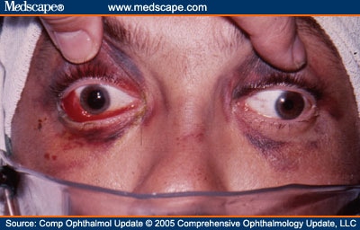
A relative afferent pupillary deficit is a necessary condition for the diagnosis of TON in unilateral cases (Figure 2). The relative afferent papillary defect is elicited with the swinging flashlight test. Light that shines into a normal eye stimulates the pupil of that eye to constrict and also stimulates the pupil of the other eye to constrict consensually. There is less pupillomotor stimulation reaching the brainstem when the light shines into the eye with optic nerve injury compared to the uninjured side, so the pupillary response is diminished. This relative afferent pupillary deficit is the basis for the swinging flashlight test.[46] A flashlight is used to illuminate one eye and after a few seconds is quickly directed into the other eye. This process is repeated until the pupillary response is assessed. If both optic nerves are normal, the pupillomotor stimulation remains the same and pupillary constriction in response to the light is equal. However, if the light is instead swung from the normal eye to an eye with an optic nerve injury, pupillomotor stimulation from the injured eye is less and both pupils will dilate. This test can be very useful for detecting unilateral optic nerve injury in an unresponsive patient. Deficits greater than 2.1 log units when measured with neutral density filters are predictive of poor visual prognosis.[47] A relative afferent pupillary defect may not be present if TON is present bilaterally.

An obvious pupillary abnormality following midface trauma. (© Regents of the University of California, reprinted with permission)
A complete and thorough examination of the eye and ocular adnexa is essential following trauma. Palpation of the orbital rim can identify step-off fractures. Periorbital swelling may mask the presence of proptosis. Tonometry and palpation should be used to rapidly identify a tense orbit from orbital hemorrhage (Figure 3). Evidence of a penetrating ocular injury should be sought. A dilated fundus examination may need to be deferred until the patient is neurologically stable. Frank papilledema in the setting of raised intracranial pressure has been documented in association with TON.[48] Often patients have evidence of other ocular pathology. Making a diagnosis of TON in this setting requires a judgement regarding the severity the ocular pathology present and whether it accounts for all the visual loss.

Orbital hemorrhage contributing to massive proptosis following orbital trauma. (© Regents of the University of California, reprinted with permission)
In addition to the ocular the examination, the visual evoked potential is useful in the unresponsive patient suspected of TON.[49,50] This is especially true in possible bilateral cases where a relative afferent pupillary deficit may not be evident. It has been reported that the visual evoked potential is useful only when it is unrecordable; in which case visual recovery is unlikely.[51] However, a recent study of unilateral TON suggests that a flash visual evoked potential with an amplitude that is within 50% of the normal side may be predictive of a favorable visual outcome.[50]
Computed tomography (CT) scanning in the setting of TON may reveal specific pathology compromising the optic nerve, including optic nerve sheath hematoma, presumed arachnoid cyst, fractures involving the greater or lesser wing of the sphenoid (Figure 4), subperiosteal hematoma (Figure 5), hemorrhage affecting the orbital apex, ethmoid or sphenoid sinus, and pneumoce phalus.[52,53] While CT scanning is clearly superior to magnetic resonance imaging (MRI) in delineating fractures of bone, MRI is superior to CT scanning for soft tissue. Often both CT and MRI are required to evaluate a given clinical situation.[54] Magnetic resonance imaging should be deferred until a metallic orbital or intraocular foreign body has been ruled out by CT scan or conventional x-ray. The reported incidence of optic canal fracture varies from 5% in the series reported by Tandon and coworkers to 92% in the series reported by Fukado.[42,55] However, the later series, as will be discussed later, lacks credibility and should be discounted. Finally, CT scanning is critical for surgical planning if optic canal decompression is contemplated.

Orbital fracture involving the greater wing of the sphenoid in association with optic nerve trauma. (© Regents of the University of California, reprinted with permission)

Subperiosteal hemorrhage with visual compromise following trauma. (© Regents of the University of California, reprinted with permission)
Compr Ophthalmol Update. 2005;6(1):11-21. © 2005 Comprehensive Ophthalmology Update, LLC
Cite this: Traumatic Optic Neuropathy: A Critical Update - Medscape - Jan 01, 2005.


Comments