Meibomian Gland Dropout
Meibomian gland dropout (MGDo) refers to the partial or total loss of acinar tissue detected by meiboscopy, meibography, or confocal microscopy.[109] To date, it is uncertain whether MGDo can result in deficiency in meibomian gland expressibility or alteration in lipid secretions that are frequently associated with lid margin inflammation commonly seen in MGD.[110,111] Although alterations to meibomian gland morphology may be suggestive of MGD, such changes are difficult to ascertain in a routine eye examination. Researchers have designed imaging systems to facilitate meibomian gland examination and diagnosis, including a combined system of infrared photography and transillumination biomicroscopy.[112] More recently, noncontact, patient-friendly meibographic techniques using infrared illumination systems have been introduced, which allow for quick and thorough examinations of morphologic changes in meibomian glands.[113,114] Using the new technology, the authors reported that CL wear accelerates age-related changes in the meibomian glands.[115] They also found that CL wearers with an average age of 32 years demonstrated an average meiboscore similar to that observed in a 60- to 90-year-old age group from the normal population.[115] Additionally, loss of meibomian glands depends on the duration of CL wear, but not on the CL materials (e.g., GP vs. conventional CLs).[115] It is unclear whether MGDo can be recovered after CL cessation.
Evidence for these meibomian gland morphologic changes was further provided by a recent in vivo histopathologic study using a laser scanning confocal microscope (LSCM).[116] This LSCM study of meibomian glands showed significantly decreased basal epithelial cell density, lower acinar unit diameters, higher glandular orifice diameters, and greater inhomogeneity of the periglandular interstices in CL wearers compared with that in controls. These authors interpret such morphologic changes as signs of MGDo, duct obstruction, and glandular inflammation. More investigation is warranted to better understand the relationship between morphologic changes of meibomian glands and MGDo.
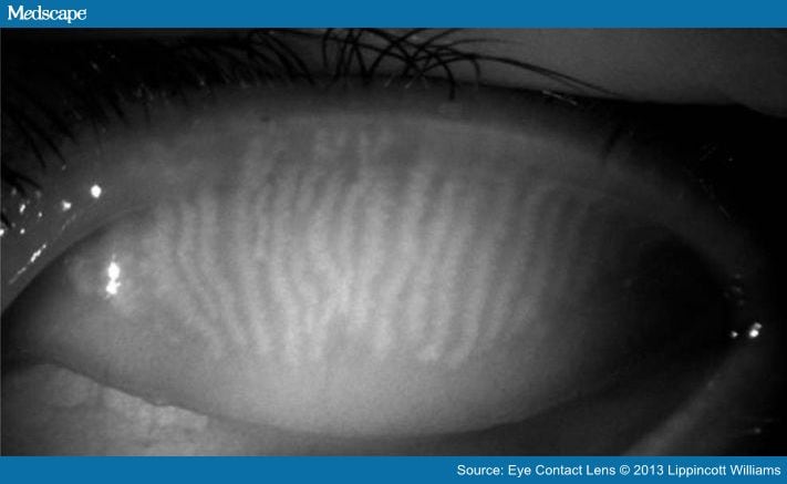
The mechanism for CL-induced MGDo is unclear. One recent study reported shortening of the meibomian glands in CL wearers (Fig. 11), particularly at the distal end, producing significantly greater effects in the upper than in the lower eyelid, supporting a mechanical theory.[115] However, other authors reported no association between MGDo in the lower eyelid and CL wear.[117] The disagreement in study results is possibly because of the authors from the latter study not examining or imaging the full extent of both upper and lower eyelids, but rather only the central portion of the lower eyelid.

Figure 11.
Meibomian gland dropout seen with noncontact infrared meibography (image courtesy of Reiko Arita, M.D., Ph.D.).
Chronic blockage of meibomian glands may eventually lead to anatomical changes in meibomian glands. One proposed theory speculates that flexing of ultrathin hydrogel lenses results in trauma to the meibomian orifices and deposition of keratic materials into the gland, impeding secretion of these glands.[118] In more recent literature, others also postulated that mechanical trauma from CLs causes hyperkeratization of meibomian glands, leading to duct blockage.[119]
Much remains unknown about the effect of CLs on meibomian glands. If flexing of ultrathin hydrogel lenses is to be blamed, we may assume that GP lenses should induce a different rate of MGDos, and yet one study did not find statistically significant differences in MGDo between GP and conventional CLs.[115] Further investigation with modern technology is needed to elucidate the mechanisms responsible for meibomian gland morphologic changes because of CL wear, to examine whether these changes differ between conventional and SiHy CLs or between different wearing modalities, and to determine how these anatomical changes affect the function (e.g., gland expressibility, lipid compositions) of meibomian glands and lens-wearing comfort.
Eye Contact Lens. 2013;39(1):115-124. © 2013 Lippincott Williams & Wilkins