Jul 8 2020
According to Penn State scientists, a special nanoparticle used to provide a localized cancer treatment suppresses the growth of tumors.
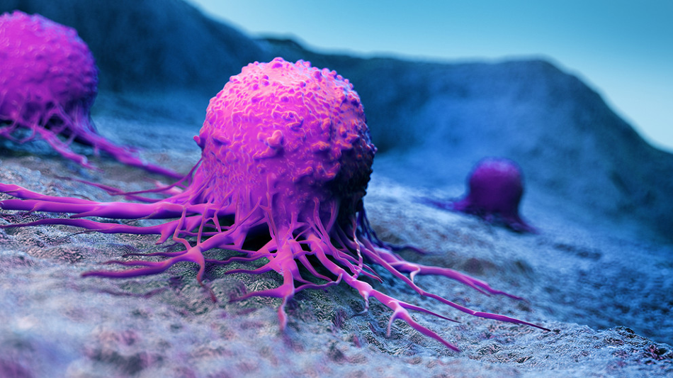 A team of Penn State researchers is collaborating on a potential new method to treat cancer by delivering a unique nanoparticle to a localized cancerous area in mice and activating the treatment through light exposure. Image Credit: Adobe Stock.
Created by Daniel Hayes, associate professor of biomedical engineering, the nanoparticles have a certain chemistry that enables a microRNA, or miRNA, to bind to it.
When a miRNA, a kind of molecule, is combined to a messenger RNA, or mRNA, it inhibits the operation of the latter. In this example, the miRNA prevents the mRNA in a cancer cell from producing proteins, which are crucial for the survival of that specific cancer cell.
In the latest study, the team used an IV to deliver nanoparticles to cancer cells present in mice. After these nanoparticles accumulated in the cancerous region, a certain wavelength of light was used to isolate the miRNA from the nanoparticles.
The miRNA subsequently binds with an mRNA present in the cancer cell and prevents it from making proteins. After that, the cancer cell dies. The study was published in the Biomaterials journal on June 22nd, 2020.
This delivery method gives you temporal and spatial specificity. Instead of having systemic delivery of a miRNA and the associated side effects, you are able to deliver the miRNA to a specific area of tissue at a specific time by exposing it to light.”
Adam Glick, Professor of Molecular Toxicology and Carcinogenesis, Penn State
According to Hayes, having spatial and temporal specificity is significant when working with cancer therapies.
“miRNA can have vastly different effects in different types of tissue which can lead to unwanted side effects and toxicity,” stated Hayes. “Delivering and activating miRNA only at the site of the tumor reduces these side effects and can increase the overall effectiveness of the treatment.”
Yiming Liu, a biomedical engineering graduate student in the Hayes Laboratory, used this technique and successfully demonstrated that skin tumors in around 20 mice that received the miRNA-coupled nanoparticle and subjected to light regressed completely within a period of 24 to 48 hours and did not grow again.
Moreover, the particular miRNA that is being used by Hayes and Glick could be more effective in destroying cancer cells when compared to other analogous techniques.
What is different about this as a therapeutic is that the miRNA that we are using can regulate a broad set of genes and is particularly powerful to treat a heterogenous disease such as cancer,”
Yiming Liu, Biomedical Engineering Graduate Student, Penn State
This could imply that the overall effectiveness of destroying a cancer cell is higher since the treatment tends to attack several points in that cell. This could result in the reduction of the ability of a cancer cell to turn resistant to the treatment, since the miRNA can pair with various mRNAs in the cancer cell, widening the means through which it can inhibit the cell from synthesizing proteins.
Cancers in the gastrointestinal system, the skin, or the oral cavity—more generally, areas that can be exposed to light using a fiber optic cable—are the cancer types that might be responsive to treatment of this kind.
We would like to develop this further for internal tumors that are more significant in terms of mortality, such as esophageal cancer.”
Adam Glick, Professor of Molecular Toxicology and Carcinogenesis, Penn State
Jacob Bailey, a student in Penn State’s Department of Veterinary and Biomedical Sciences, and Mohammad Abu-Laban, Shue Li, and Cong Chen, students in Penn State’s Department of Biomedical Engineering, are the other authors of the study.
This study was financially supported by the Human Health and the Environment Seed Grant program, funded by the Penn State College of Medicine, Penn State Cancer Institute, Huck Institutes of the Life Sciences, Social Science Research Institute, Clinical and Translational Science Institute, Materials Research Institute, Institute for Computational and Data Sciences, and Institutes of Energy and the Environment.
Journal Reference:
Liu, Y., et al. (2020) Photocontrolled miR-148b nanoparticles cause apoptosis, inflammation and regression of Ras induced epidermal squamous cell carcinomas in mice. Biomaterials. doi.org/10.1016/j.biomaterials.2020.120212.
Source: https://www.psu.edu