Biology & Chemistry of Arrays
Types of Assays
The most common method of detecting proteins in a solution is a sandwich assay, which is the ELISA. The sandwich assay involves the formation of a three-layered structure, where capture probes and detection probes form a sandwich around a protein, specifically the analyte of interest, as shown in Figure 1. The capture probes are preferably antibodies but could be aptamers,[23,24] diabodies[25] or Fab fragments,[26,27] for example. For autoantibody detection, the capture probes can be autoproteins or peptides, and the detection probes are anti-species antibodies, so that they form a sandwich around the autoantibody.[28,29] The capture antibody is directly immobilized on the surface of a solid (Figure 1A). To have a highly specific assay, a monoclonal antibody is preferably used, and it will bind to a certain type of binding site, called an epitope, on a protein. If polyclonal antibodies that bind to multiple epitopes are used as capture probes, other highly abundant proteins that share the same epitope might bind to the antibodies and prevent the target protein from binding, which in turn hampers detection of the target protein. Normally, polyclonal antibodies can have a higher probability of nonspecific binding if multiplexing measurements are performed, where multiple probes and analytes are involved.

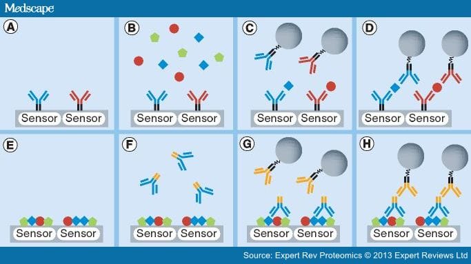
Figure 1.
Schematics of sandwich and reverse-phase assays. (A) The sensors are immobilized with capture probes. In this case, antibodies are selected as capture probes. (B) After blocking the rest of the surface, the sample containing target proteins is introduced and incubated. The complementary pairs of antibody and its antigen are displayed in the same color. Thus, irrelevant proteins shown as green pentagons are not captured by capture probes. (C) After washing, the detection probes are added. They can be preconjugated with tags (which are shown as gray circles) or additional steps can be performed to attach the tags to them. (D) If the detection probes do not crossreact with other proteins or capture probes, the final assembly becomes a three-layered structure of sandwich assays. (E) In the reverse-phase assays, the samples containing target proteins are spotted on the sensors. (F) After neutralizing the surface, the primary antibodies are added and incubated. These antibodies can recognize their target proteins, which are shown as blue squares. (G) After washing unbound primary antibodies, the secondary antibodies with tags are introduced. These secondary antibodies bind to the other parts of the primary antibodies, which are indicated in orange. (H) The final assembly of reverse-phase assays.
The second antibody, known as the detection probe, is delivered in solution after the protein of interest is incubated and captured by the capture probes. It binds to a second epitope on the captured protein and offers additional specificity in conjunction with the capture probes. If there is another protein that has the first epitope that binds to the capture probes but not the second epitope, this protein will not bind the detection probes. This is a major advantage that the sandwich assays have over other direct assays, where only one type of antigen-specific antibody is used. In addition, the detection probes are premodified with a reactive chemistry, enabling easy attachment of the probes to the tag of interest, which converts binding events into readable signals. In the ELISA, the tag of interest is enzymatic or fluorescent, while it is magnetic when using magnetic immunoassays. Sometimes, the detection probes are directly conjugated with the tag of interest, but normally the probes are biotinylated and the tag of interest is conjugated with avidin or streptavidin to bind them together using biotin–avidin interaction, which is one of the strongest forms of noncovalent binding.
Another type of assay widely used is a reverse-phase assay, where many patients' blood samples are analyzed simultaneously.[20,30] In the reverse-phase assay, patient samples containing proteins of interest are immobilized on the surface instead of attaching capture probes to the surface (Figure 1E). Then, a solution containing primary antibodies specific to the protein of interest is introduced (Figure 1F), and these will bind to the immobilized protein of interest on the surface. Subsequently, a secondary antibody, which is conjugated with the tag of interest or biotinylated, is delivered to the surface, and will bind to the primary antibodies already bound to the protein of interest. Usually, the patient samples are a mixture of numerous kinds of proteins if not purified. When they are immobilized on the surface, all proteins bind to the surface competitively if no specific attachment method is applied. For the protein that has a relatively low concentration, the number of bound proteins is lower than other proteins, and this provides the primary antibodies with fewer binding sites. As a result, a reverse-phase assay has relatively low sensitivity compared with sandwich assays.
Protein Immobilization
The capture probes, or in the case of the reverse-phase assay, the proteins of interest in a sample can be immobilized on the surface via a variety of different methods. Ideally, the probes will be attached to the surface without any conformational changes and with their activity intact. One of the methods is nonspecific physical absorption where electrostatic forces between the probes and thin film polymer on the surface dominate.[31,32] For instance, a cationic polymer can be deposited on the surface to attract negatively charged proteins or antibodies.[33,34] Poly-l-lysine has been widely used in protein microarrays.[35] It has also been reported that polymers such as polyethyleneimine provide both an easy and effective means of physical absorption-based antibody binding.[36,37]
Another method is to employ covalent binding chemistry where new molecular bonds are formed.[31,35] This method of immobilization is typically more effective and reproducible than physical absorption because only certain reactive groups on the probes and a layer on the surface are involved to form new bonds. A common reactive group of the proteins is primary amines on lysines and arginines. Essentially, all proteins can be immobilized by amine-reactive chemistry that provides stable amide bonds. N-hydroxysuccinimide (NHS) and 1-ethyl-3-(3-dimethylaminopropyl)carbodiimide (EDC) conjugation is the most common amine-reactive chemistry. The EDC reacts with a carboxyl group on a layer of the surface, forming an amine-reactive intermediate. This intermediate can react with an amine group on the proteins to form an amide bond, but it is unstable and short-lived in aqueous solution due to hydrolysis. The addition of NHS stabilizes the intermediate by converting it to an amine-reactive NHS ester, which increases the efficiency of amino-reactive chemistry mediated by the EDC.[38]N-hydroxysulfosuccinimide is a negatively charged analog of NHS and can be an alternative choice for stabilizing the intermediate activated by EDC.[39]
Alternatively, thiol–gold conjugation can be utilized to attach the probes with thiol groups to the gold surface.[40,41] Since all antibodies contain interchain disulfide bonds, it is possible to reduce these bonds selectively by using reducing agents, such as 2-mercaptoethylamine or dithiothreitol, to make thiol groups.[42,43] This method will create a portion of half antibodies because the disulfide bonds can be broken. The half antibodies can be separated from a mixture of antibodies by column chromatography. If this kind of antibody is not desired in an experiment, 2-iminothiolane, also known as Traut's reagent, can be used to create a free sulfhydryl group by reacting with amine groups on antibodies.[44,45] Once sulfhydryl groups are generated, the target surface, an area where the antibodies are to be attached, is coated with gold and the antibodies with sulfhydryl groups can be immobilized on the surface.
Surface Blocking
After immobilizing the capture probes on the surface of arrays, the remaining reactive area must be neutralized or blocked to prevent any future nonspecific binding of proteins, detection probes or tags.[46] Otherwise, nonspecific binding elevates the background signal such that readout signals can be overestimated. It is difficult to subtract these nonspecific signals from the readout signals because they are not necessarily constant within an array or between arrays. Another problem that is encountered is that sensitivity at a low concentration of the analyte can be impaired, as the analyte can be captured outside of the sensing area and become depleted in the reaction well. To get reproducible and reliable signals, the remaining reactive surface is treated with highly abundant proteins such as albumin or casein. The most common blocking reagent is bovine serum albumin (BSA) at a range of concentration from 1 to 3%.[47] Nonfat milk blocking buffers have been used to block the surface, but BSA is usually more effective than nonfat milk for biotin–avidin systems because it contains a single purified protein.[48] Generally, a blocking solution with fewer kinds of proteins has a lower chance of cross-reactivity. The authors observed that BSA blocks well in GMR sensors, and used the sensors coated with BSA as negative control sensors.
Expert Rev Proteomics. 2013;10(1):65-75. © 2013 Expert Reviews Ltd.