Three-dimensional imaging just got faster and far simpler, thanks to scientists at Johns Hopkins University (JHU; Rockville, MD) and Ben-Gurion University of the Negev (BGU; Beer-Sheva, Israel), with their recent demonstration of the FINCHSCOPE—a motionless microscopy system based on Fresnel incoherent correlation holography (FINCH)—and its use in recording high-resolution 3-D fluorescent images of biological specimens.
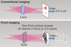
Gary Brooker, director of JHU’s Microscopy Center, and Joseph Rosen, professor of electrical and computer engineering at BGU, first described their method of recording holograms, known as “FINCH,” last year.1 FINCH is basically a single-beam holographic system that works without a laser, as opposed to a conventional dual-beam holographic interference system that in the most classical sense uses the interference of a split laser beam. In conventional imaging, only one plane is in focus at any time and multiple images at different planes of focus need to be captured to obtain all of the information in a 3-D image. In contrast, the FINCH technique captures all of the 3-D information in one hologram such that the 3-D information is encoded in a series of Fresnel zone plates (see figure).
The primary benefit of FINCH is that it can produce a hologram with incoherent light. There’s no need to illuminate the sample with a laser to get a hologram and a 3-D image. It’s essentially a 3-D camera, according to Brooker and Rosen. There are no moving parts and this technique doesn’t require complicated alignment. Normally you’d have to change the position of the lens or the sample many, many times to get individual image planes in focus.
Conventional 3-D imaging involves acquiring multiple images on multiple planes and then reconstructing the images. The FINCH concept, instead, relies on microscope objectives with the highest resolving power, a spatial-light modulator, a charge-coupled-device (CCD) camera, and some simple filters to enable the acquisition of 3-D microscopic images without scanning multiple planes.
The scientists illuminate the sample with a lamp that is not coherent, so it is illuminated just like in any normal fluorescence microscope through epifluorescence, and the emitted light bounces off the spatial light modulator. The phase of the emitted signal gets altered and interference occurs between two beams that are coincident with one another upon the camera sensor (the CCD camera). This creates a hologram that can then be processed to create individual image planes or a 3-D image.
Epifluorescence microscope
Moving their original concept forward, the scientists applied the FINCH technology to epifluorescence microscopy in the “FINCHSCOPE,” and ran a series of demonstrations on fluorescent beads, fluorescently labeled pollen grains, autofluorescent Convallaria rhizom, and fluorescently labeled nerve fibers in a skin section.2 Rosen and Brooker report that the FINCHSCOPE was able to rapidly create high-resolution images of microscopic specimens with each plane in focus, without sectioning or the need for movement in the z-direction or any other movement of the microscope or specimen. The resulting holograms revealed fluorescent specimens in focus at all planes in the image space, almost as if the images had been taken with a standard microscope by changing the focus to obtain each image section.
Rosen notes that while each reconstructed section is currently not completely confocal, 3-D reconstructions free of blur could be created by deconvolution of the holographic sections as is typically achieved in widefield microscopy.
Brooker and Rosen also observed that their FINCH technology is 25 times faster than wide-field or confocal microscopy, largely because everything is in focus and no scanning or sectioning is involved. “Take one snapshot and you’ve got that volume at that timeframe. Take the next snap, and over and over again. The potential to track objects moving rapidly in 3-D space is very real,” says Brooker.
Many imaging applications should be amenable to FINCH technology, according to Rosen. It shows tremendous potential for wave-based applications ranging from endoscopy, ophthalmology, CT scans, x-ray imaging, and ultrasounds, to homeland security, 3-D photography, and 3-D video.
CellOptic, a company cofounded by Rosen and Brooker, owns the FINCH technology and is supporting its commercialization.
REFERENCE
1. J. Rosen and G. Brooker, Optics Lett. 32, 8 (April 15, 2007).
2. J. Rosen and G. Brooker, Nature Photonics, DOI: 10.1038/nphoton.2007.300.

