
Array-Based Cancer Diagnostics
Cell Surface Sensing
Genotypic and phenotypic differences in cells result in different levels of polysaccharides, proteins and lipids on the cell surface.[21,22] This cell surface signature was used to differentiate cell genotype. Rotello and coworkers used an array of gold NPs (AuNPs) with varying headgroups to differentiate the cell surfaces. This strategy employed the strong quenching ability of AuNPs[23] to enable the use of fluorescent displacement assays, where displacement of a quenched fluorophore from the recognition element signals the binding event.[24]
In an earlier study, Rotello et al. developed an array of fluorescent polymer-conjugated AuNPs to detect different cancer cell types. Cationic AuNPs were used as recognition elements to noncovalently bind negatively charged poly(p-phenylene-ethynylene) polymer (PPE-CO2) as the transducer.[25] The electrostatic interaction between PPE-CO2 and AuNPs quenched the fluorescence of the polymers, which was recovered to varying degrees upon incubation with cells. Distinct and differentiated patterns were observed with human cancerous (MCF-7), metastatic (MDA-MB-231) and normal (MCF10A) breast cell lines. These cell lines, however, came from different individuals, making it possible that differentiation was based on individual genetic background. To avoid individual variation, isogenic cell lines derived from BALB/c mice (CDBgeo [normal], TD [cancerous], and V14 [metastatic]) were used to demonstrate the ability of the sensor array. In a further study, the author demonstrated that green fluorescent protein (GFP) could also be used as a transducer with high quantum yield, low aggregation and high sensitivity. This GFP–AuNP array sensor was able to discern different cancer cell types with as low as a 5000 cells (Figure 2) lower limit of detection, compared with the 20,000 cells used in AuNP–PPE-CO2.[26] Recently, quantum dots were used in combination with AuNPs to provide a sensor array.[27]

Figure 2.
Mechanism of detecting cancer cell types using green fluorescent protein–gold nanoparticle sensor array. (A) Structures of the NPs with different head groups R used as an array of recognition elements in the sensor. (B) Structure of GFP used as the transducer of the sensor array. (C) Schematic illustration of GFP–NP complexes for cell surface sensing. The fluorescence from GFP is turned off once GFP–NP complexes are formed due to the strong quenching property of gold NPs. Due to competitive binding between cells and GFP to the NPs, different amount of GFP are being released in the presence of different cell lines based on their binding affinities.
GFP: Green fluorescent protein; MW: Molecular weight; NP: Nanoparticle; pl: Isolectric point.
Adapted with permission from [26].
Biomolecules, such as antibodies and/or synthetic aptamers, are typically used for specific recognition. They can, however, also be used for selective recognition.[28] Nonspecific aptamers were used to protect bare AuNPs from salt-induced aggregation. Upon addition of target cells, the competition between citrate-capped AuNPs and the cells will detach various amounts of aptamers from the AuNPs. Depending on the target cell line, different aggregation levels generated color change patterns that were used to differentiate cell lines. Through the use of two thrombin aptamers (Tro-1 and Tro-2) and one human IgE aptamer (HIgE-1), human cancer cells (Junkat, Reh and Raji) and normal human cells (WIL2-S) were differentiated.[28]
Aptamer-conjugated magnetic NPs (ACMNPs) were developed for cancer cell sensing using magnetic signal transduction.[9] This magnetic NP-based sensor was fabricated by conjugating streptavidin-coated iron oxide NPs with biotin-labeled aptamers. ACMNPs bind to the target cells through specific and nonspecific interactions between the aptamer ligands and the membrane receptors. The spin–spin relaxation time (T2) read out was measured by nuclear magnetic resonance spectroscopy. Using the magnetic property of their NPs, the authors were able to detect ten cancer cells in buffer and 100 cancer cells in other biological complex media, such as fetal bovine serum, plasma and whole blood.
Magnetic glyco-NPs were also used for array-based sensing. In this approach, a variety of carbohydrates were coated as ligands on magnetic NPs to explore the interactions of carbohydrate–cell membrane receptor upon cell incubation (Figure 3).[29] Similarly with the ACMNPs, different degrees of aggregation upon incubation with cancer cells generated different local magnetic field gradients and altered the transverse relaxation time. Magnetic glyco-NPs were able to differentiate several cancer cell types with a sensitivity of 105 cells/ml (Figure 4).

Figure 3.
Structures of magnetic glyconanoparticles with different carbohydrate ligands.
MGNP: Magnetic glyconanoparticle.
Data taken from [29].

Figure 4.
Plot generated from linear discriminant analysis plot for ten cell line differentiation using magnetic glyconanoparticle sensor array. 3D linear discriminant analysis plot of ΔT2 patterns generated from the magnetic glyconanoparticle array after different cell line incubations (105 cells/ml). ΔT2 patterns of different cell lines were reduced into simpler components, linear discriminants (LD1, LD2 and LD3). Full differentiation of the ten cell lines was achieved.
LD: Linear discriminant.
Adapted with permission from [29].
Cancer-cell sensing using array-based sensors provides a proof of concept for rapid cancer diagnosis. However, moving from cancer cell sensing in vitro to real cancer cells from patients will need extensive development to transform the methodology into a useful and meaningful clinical screening tool.
Tissue Biopsy Sensing
The above studies demonstrate the use of NP array-based sensing for cell surfaces in cell suspension. Solid tissues are much more complex systems, making cell surface sensing impractical for identification of solid tumors. Currently, excisional biopsy is the standard approach for cancer diagnosis with solid tumors. In this method, a tissue specimen is obtained from the patient to be examined by a pathologist. Although this method detects meaningful phenotypic differences, it is very time consuming, low throughput and requires high expertise.
As a potential alternative for point-of-care diagnosis, chemical nose sensors have been applied to tissue sensing. Given the complexity of tissues, Rotello and coworkers have focused on sensing of intracellular proteins using an array of GFP–AuNP conjugates. Protein sensing approaches using chemical nose sensors have been developed, facilitating this strategy.[13,28,30] In their studies, a mouse metastasis model was generated by inoculating NCI-H1299 non-small-cell lung cancer cells that metastasize to multiple organs. Tissue lysates were prepared from tumor tissue microbiopsies (˜1000 cells) using lysis buffer that contained protease inhibitors. By focusing on unique proteomic profiles, GFP–AuNP sensors were able to detect site-specific metastatic tissues from healthy ones with high sensitivity, providing a potentially rapid and effective tool for clinical diagnostics.[31]
Urine Samples
Many cancer diagnoses are accomplished through excisional biopsy. Despite the usefulness of this method, these techniques are unpleasant for patients. Therefore, efforts have been made to achieve sensing methodologies using noninvasive biofluids. Noninvasive biofluids are easily-accessibe biological samples, which usually do not involve instrument insertion into the patient's body. Besides breath, urine, saliva and sweat, other minimally invasive procedures such as nipple aspiration along with ductal lavage can provide many cells collected from the milk ducts.[32]
Currently, NP array-based sensors have focused on the use of urine[10] and breath[33] as samples in cancer diagnosis. Although urine is an attractive noninvasive biofluid for analysis, concentrations of naturally occurring biomarkers are typically low. To compensate for this limitation, an array of sensors with the ability to amplify biomarker concentration in urine has been developed.[9] For this purpose, a class of engineered mass-encoded peptides with specific protease-sensitive moieties conjugated to iron oxide NPs were synthesized. These synthetic biomarkers passively accumulate in the cancer tissue after administration. Aberrantly active proteases in the tumor subsequently cleave the protease-sensitive agents of these NPs, with the resulting fragments excreted in the urine (Figure 5).[34] Using a library of different substrates as the protease-specific mass signatures, differentiation between different proteases was possible. The unique profiles of the isobar-coded reporters for each protease were determined by Pearson's correlation analysis.

Figure 5.
Use of synthetic biomarker-conjugated nanoparticles for urinary monitoring. (A) Synthetic biomarker library conjugated onto nanoparticles (NPs). (B) Accumulation of NPs in disease tissues. Cleavage of the mass-encoded peptides from NPs by active proteases allows their filtration into the urine. (C) Detection of biomarker peptides in urine using LC-MS/MS.
hv: UV light irradiation; LC-MS: Liquid chromatography-mass spectrometry; MS: Mass spectrometry.
Adapted with permission from [10].
Breath Samples
Breath sensing is perhaps the least invasive of all diagnostic strategies. Metabolic reactions in the body generate different volatile organic compounds (VOCs). These VOCs include hydrocarbons, alcohols, aldehydes, ketones, esters, nitriles and aromatic compounds. VOCs can be detected in diverse biosamples, such as cancer cells, blood, urine, skin/sweat[35,36] and breath. In fact, besides cancer detection, VOCs are being used as new diagnostic-based biomarkers for detection of different diseases such as diabetes,[37] Alzheimer's, Parkinson's,[38] and chronic kidney disease.[39]
Compared with healthy individuals, cancer patients express different VOC compositions in their breath due to the different activities of cancer cells.[33] These activities can generate very subtle changes in the concentration and composition of VOCs in the blood stream. Through constant exchange between the lung and bloodstream, these subtle changes in VOC compositions can be transported to the patient's breath, creating distinct breath signatures for each cancer type.[40] In a healthy breath, the concentration of several VOCs are normally in the range of 1–20 p.p.b..[11] However, they can be detected in the levels of 10–100 p.p.b. in some cancer types. These changes in concentration and composition mixture of VOCs have made it possible to not only distinguish between the breath of healthy individuals and cancerous patients, but also differentiate between different types of cancer.[11,33] Previously, VOCs have been detected using gas sensors such as gas chromatography,[41] and ion mobility spectrometry.[42] However, the downsides to these methods are that they are time consuming and require large size, expensive instrumentation and an expert operator. Moreover, to improve the detection in some of these devices, capturing and preconcentrating the breath sample is a prerequisite step.[43,44]
Using the advantages of NPs, a nanoscale artificial nose has been designed by Haick and co-workers.[44] This simple, cost effective and portable sensor is able to detect cancer by analyzing the VOCs using pattern recognition methods. The nanoscale artificial nose is capable of identifying different odors even at very low concentrations and subtle differences.[44] The gas sensor is based on an array of highly cross-reactive chemiresistors made of AuNPs with different organic capping layers.[11] In the resulting sensor, electrical conductivity was provided by the metallic particles and the organic capping layers create sites used to capture the analytes (Figure 6).[11]
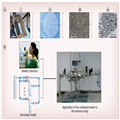
Figure 6.
Illustration of the diagnosis of lung cancer using breath testing. (A) (i) Photograph of the array of chemiresistors; (ii) Scanning electron microscopy image of a chemiresistor; (iii) Scanning electron microscopy image of gold nanoparticle film located between two adjacent electrodes; and (iv) Transmission electron microscopy image of the monolayer-capped gold nanoparticles. (B) Testing of the exhaled and simulated breaths.
MFC: Mass flow controllers.
Reproduced with permission from [11].
Due to the chemical diversity of sensor materials, each sensor of the array shows a unique response to a certain group of VOCs. This means that the characteristic signal (electrical resistance) of each sensor in the array changes specifically when exposed to a specific VOC, which could be the cancer specific odor (Figure 7).[44] Consequently, for each cancer type, a distinct fingerprint is produced from the array of cross-reactive sensors. Using this gas sensor along with pattern recognition methods, it is possible to discriminate different cancer types and stages (Figure 8).[33,45] It is also worth mentioning that these sensor arrays have detection limits of 1–5 p.p.b. or even down to approximately 10 p.p.t..[33] Breast, lung, colon, gastric, colorectal, head-and-neck and prostate cancer are the cancer types that have been detected using this sensor.[33,44] Carbon nanotube arrays have been used in a similar fashion.[46,47]

Figure 7.
Typical responses of the breath sensor. (A) Schematic representation of the gold nanoparticle sensors (not drawn to scale). Typical resistance responses of sensors functionalized with: (B) 2-ethylhexanethiol; (C) decanethiol; and (D) 2-mercaptobenzoxazole, to the breath of healthy individuals as well as patients with LC, CC, BC and PC.
BC: Breast cancer; CC: Colon cancer; LC: Lung cancer; PC: Prostate cancer.
Reproduced with permission from [33].

Figure 8.
Classification results of the sensor array's resistance response. Principle component analysis plots of the sensor responses to: (A) LC and healthy controls; (B) CC and healthy controls; (C) BC and healthy controls; (D) PC and healthy controls; and (E) all cancer patients and healthy controls together.
BC: Breast cancer; CC: Colon cancer; LC: Lung cancer; PC: Prostate cancer; PC1: Principal component 1; PC2: Principal component 2.
Reproduced with permission from [33].
It should also be mentioned that although breath sensing is a novel method for cancer detection, the approach has some limitations. First of all, there are not dramatic changes in VOCs in the early stages of cancer development, only certain stages will cause the expression of these VOCs. Second, the conditions and type of foods and drinks consumed by patients can influence results. The results can also be affected if the patients have other diseases and are using other medicines.[33] Therefore, having sufficient controls over sample collection is essential when using this type of noninvasive sample.
Nanomedicine. 2014;9(10):1487-1498. © 2014 Future Medicine Ltd.


