As 3D printing becomes more and more used in a wide range of fields, medical science is not left behind. From the more standard uses such as printing medical equipment and prosthetics to more advanced uses like printing cartilages and bones, the success of 3D printing technologies in the medical field is rapidly growing.
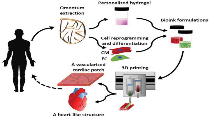
One of the last breakthrough is the world’s first 3D vascularised engineered heart using the patient’s own cells and biological materials. Until now, scientists have only been successful in printing only simple tissues without blood vessels. Researchers from Tel Aviv University used the fatty tissue from patients to separate the cellular and acellular materials and reprogrammed the cells become pluripotent stem cells. The extracellular matrix (ECM) was processed into a personalized hydrogel that served as the basis from the print.
This heart is made from human cells and patient-specific biological materials. In our process these materials serve as the bioinks, substances made of sugars and proteins that can be used for 3D printing of complex tissue models… At this stage, our 3D heart is small, the size of a rabbit’s heart, but larger human hearts require the same technology.
After being mixed with the hydrogel, the cells were efficiently differentiated to cardiac or endothelial cells to create patient-specific, immune-compatible cardiac patches with blood vessels and, subsequently, an entire heart that completely matches the immunological, cellular, biochemical and anatomical properties of the patient. The difficulty of printing full-blown organs were being tackled for a long time and we already talked about it in the past.
The development of this technology may completely solve both the problem of organ compatibility and organ rejection.


Goodbye hackaday comments. It was fun while it lasted.
My question is, could it hold together after the hydrogel is removed? If it needs to stay in the hydrogel for a while, how do they Supply nutrients to it well it’s in there?
Why bother with a single, full-sized heart? Just give the patient a dozen rabbit-sized hearts plumbed in parallel, series, or series-parallel. That way, future failures have automatic redundant backups already in place! Plus, you could freak people out with your crazy or almost non-existent pulse xD
“Sweetheart, why is your chest humming?”
“Oh, that’s just the pulses from my heart array.”
Sorry, but this paper doesn’t include anything close to an actual heart. At best, there is an upside down pear shaped blob with some hand-drawn tubes running down the side. It doesn’t have any of the actual structures found in a human heart such as valves, trabeculations, or vascular networks. It’s cute and colorful and it kinda resembles a heart, but it isn’t one. The printing method they used is nice, but the cells in the “heart” (fake news?) we’re encapsulated yet never shown to do anything other than survive printing.
I’m not a bio engineer, but as far as I understand, you are right.
The point of their research is to “create patient-specific, immune-compatible cardiac patches with blood vessels” which will lead to heart’s printing in the future. But by now it’s more a custom bloody cardiac muscle than a fully functional organ (whatever the size).
“May completely solve the problems of biocompatibility and rejection” — Yeah, but it also solves a much bigger problem… That of “Organ Availability”. Many people die every year on the transplant list (According to https://www.americantransplantfoundation.org/about-transplant/facts-and-myths/ it’s roughly 7,300 each year) due to lack of suitable organs available. The ability to 3d print 100% compatible organs without requiring the donation (often due to death) from another patient is a huge gain if we can get there. This looks to me like a giant step in the forward.
@benchMark has some valid points, but, the reality is that since the heart is printed from pluripotent stem cells, if researchers can provide the appropriate stimuli (as the article calls it “teaching the cells to behave like a heart”), the formation of those structures (valves, trabeculations) should be automatic. As I understand the article, the printed heart does include vascular networks. It would stand to reason that the vascular networks are required up front in order to be able to supply nutrition (and possibly as conduits for the necessary stimuli for training) during “training”.
@Jack Huffington The way I read the article, I don’t think the hydrogel is ever removed. I believe that over time, the cells will consume it, leaving new capillaries in its place in some cases and bonding directly to adjacent cells in others, creating the necessary muscle fibers and other structures necessary for a functioning heart. This is quite similar to how the process works in utero, but starting with a full-size collection of stem cells instead of a small blob with a lot of the structure already defined would likely provide much faster results an attempting to grow an ex-vivo heart from a small collection of stem cells. Presumably it would also help with the process of convincing the stem cells to become heart cells.
This paper’s “heart” has no vascular networks. Just a couple hand-drawn, dead-end tubes running down the side. Extruding stem cells in decellularozed ecm digest is one thing. Calling it a heart, much less a tissue without showing any degree of function is misleading to those who pick up these articles and read nothing but the headline/abstract. Granted, many of them are paywalled. Furthermore, a heart’s morphogenesis is an incredibly complex process that starts with just a tube. Printing a heart will most likely mean one of two things: printing this tube and figuring out a way to make it become a heart, or printing an actual anatomical heart from a similar ECM digest.
Why go to all the trouble of printing a heart when you can just go to the slaughterhouse and pick up a ready made one?
https://www.cleveland.com/healthfit/2012/08/ghost_heart_a_framework_for_gr.html
This aligns with the challenges of printing multiple extrusions or for living structure… cells/tissues/media. Seems an advancement in 3D printing will be more advanced heads like say color printing with printer heads versus just printing in black.
Maybe multiple streams with controlled entry into one extrusion/print point is a way too.
Also, what is great about this method is it looks like is an autograph method. This in my opinion is critical in integrating into the body and not having to deal with the variables of rejection that must be considered with other non-autograph methods.
This has to be the future direction method of thinking in my opinion. Especially for those that want to teleport. How else can someone be ablated and then re-assembled next to real time? A little advancement in super glue and healing time… I mean duh?
Seriously, though… this type of research is in the correct direction. We need to advocate more of the research like this say like the tooth transplant research that looks promising so we have a more autograph or at least bio-equivalent future.
Sickle cell anemia is being cured and looks like AIDS in the same autograph… though with genetic engineering ways.
https://www.youtube.com/watch?v=qNxwq10kXXk
Excellent post! Can’t get any better than this. Thanks!