In a recent study published in the journal Science Robotics, a team of researchers created a magnetic microrobot probe that can quantify the stiffness and traction force of tumor cell colonies and developing vertebrate embryos.
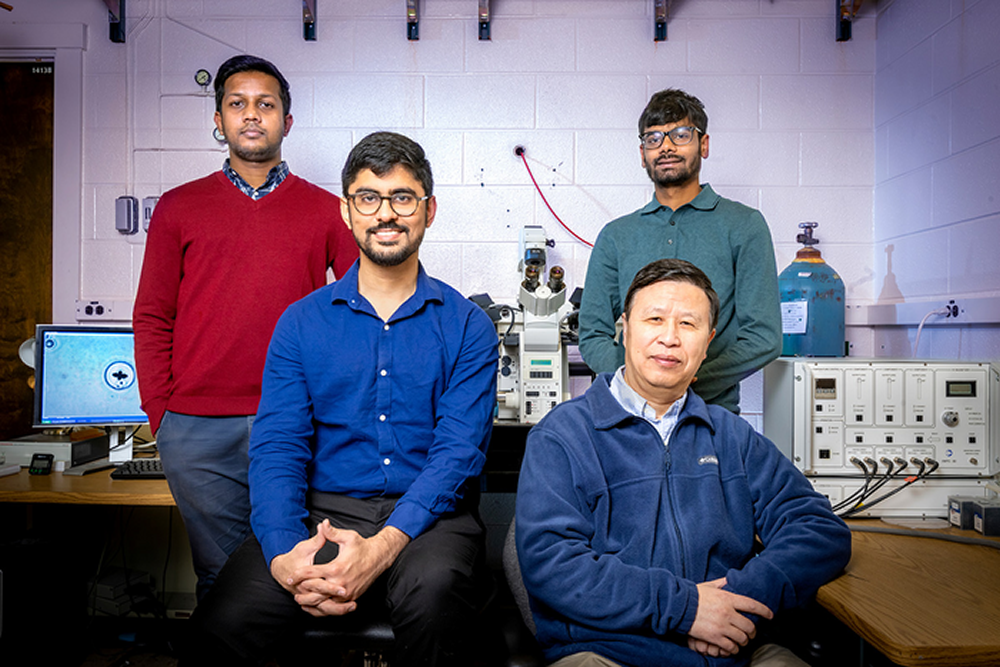
Professor Ning Wang, front right, is joined by researchers from left, Fazlur Rashid, Kshitij Amar, and Parth Bhala. Image Credit: Fred Zwicky
Background
A growing body of evidence suggests that the two fundamental mechanical parameters in the functioning of cells and tissues and their role in development, physiology, and a wide range of diseases are traction forces or cell-generated contractile forces and stiffness. However, the actomyosin-dependent traction forces are stronger in the monolayer epithelia, while stiffness is dominant in the multilayer epithelia.
The quantification of both stiffness and three-dimensional (3D) traction forces at the same location of biological tissue has proved to be challenging due to the nature of the probe required to measure each of these parameters. Quantifying traction forces requires a soft probe, where the deformation of the probe indicates the traction force. Stiffness is quantified using a hard probe that can apply stress or torque force to deform the sample. While various probes have been developed to quantify stiffness and 3D traction forces, quantifying the two parameters in vivo using the same probe continues to be challenging.
About the study
In the present study, the researchers developed a remotely and magnetically controlled microrobot probe made of ferromagnetic cobalt-platinum microcrosses that were biocompatible. Each microcross was embedded inside a stiff poly (ethylene glycol) (PEG) microgel with an arginine–glycine–aspartic acid conjugate.
The stiff microcross-embedded microgel could be rotated using an oscillatory magnetic field and was used to quantify the stiffness. The 3D traction forces were then quantified by exposing the PEG microgel with embedded the fluorescent nanoparticle to ultraviolet light to photodegrade and soften the probe.
The probe was used to quantify the stiffness and 3D traction forces of cell colonies of malignant tumor-repopulating cells isolated from mouse melanoma cell lines grown on a fibrin gel matrix. Once the stiffness of the cancer cell colony was measured, the PEG gel was softened by exposing the microrobot to ultraviolet light and quantified the deformation of the gel by the surrounding cancer cells.
Furthermore, the microrobot probes were also used to measure the 3D traction forces and stiffness of Zebrafish and mouse embryos.
Results
The results demonstrated that based on the elasticity of the 3D substrate, the modulus or stiffness of the malignant tumor cell colonies changes, but the 3D traction forces do not vary. Furthermore, the simultaneous analysis of the stiffness and 3D traction forces at the same site of the cell colony shows no correlation.
The quantification of stiffness and traction forces in Zebrafish embryos revealed substantial oscillations in traction forces during embryonic development, with shear and normal traction forces of comparable magnitudes. In contrast, mouse embryos also indicated oscillations in traction forces during development, but the normal traction forces were higher than the shear traction forces. During the blastocyst stage, mouse embryos generated larger oscillations in compressive and tensile traction forces than in shear traction force.
Compared to previous methods using magnetic probes where the dynamic properties of biological samples could not be measured, the researchers reported that the microrobot magnetic probe could quantify various rheological properties, including loss modulus and storage modulus. Furthermore, the method of softening the microrobot probe using ultraviolet light allows a range of 3D shear and normal traction forces of biological samples to be quantified.
The possible alteration of measured traction forces due to the magnetic microcross itself was examined, and the results indicated that the microcross had a negligible effect on the 3D traction forces. Moreover, the shear and normal traction forces did not change with the softening or swelling of the microgel due to ultraviolet light exposure. Additionally, the orientation and position of the microcross inside the PEG microgel relative to the magnetic field did not influence the measurements of stiffness. Furthermore, the metallic microcross did not interfere with the softening of the PEG microgel when exposed to ultraviolet light
Conclusions
To summarize, in the present study, researchers developed a remotely controlled magnetic microrobot to quantify the 3D traction forces and stiffness of cell colonies in vivo at the same time. The microrobot was tested on cell cultures of malignant tumor cell colonies, as well as on Zebrafish and mouse embryos. The results indicated that the microrobot could successfully measure the stiffness and traction forces of cell colonies in vivo and that traction forces play a major role in early embryonic development regulation. This microrobot probe could be used to study the mechanoregulation of embryos and cell cultures.
Journal reference:
- Mohagheghian, E., Luo, J., Yavitt, F. M., Wei, F., Bhala, P., Amar, K., Rashid, F., Wang, Y., Liu, X., Ji, C., Chen, J., Arnold, D. P., Liu, Z., Anseth, K. S., & Wang, N. (2023). Quantifying stiffness and forces of tumor colonies and embryos using a magnetic microrobot. Science Robotics, 8(74). https://doi.org/10.1126/scirobotics.adc9800, https://www.science.org/doi/10.1126/scirobotics.adc9800
 Mitochondrial fusion critical for adult neurogenesis and brain circuit refinement