The Asylum Research MFP-3D-BIOTM is setting the bar for combining AFM and optical microscopy in bioscience research. It is the only bioAFM that smoothly integrates with a wide variety of optical methods while maintaining uncompromised AFM image resolution, force measurement performance, or application adaptability. This, combined with unequaled assistance from Asylum specialists, makes it the finest, most productive bioAFM to help researchers accomplish their research goals.
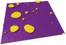
2.5 µm scan of supported lipid bilayers (5 nm tall) adsorbed onto mica and imaged in buffer. Image Credit: Asylum Research - An Oxford Instruments Company
The performance and versatility to get the results needed
- Simple, liquid imaging with great resolution for delicate biological samples
- Performance management in force mechanics and force spectroscopy
- Integrating optical microscopy with AFM performance that is uncompromised
- Unparalleled assistance from bioAFM specialists to achieve the desired outcomes
- Unmatched adaptability to meet the various demands of various consumers
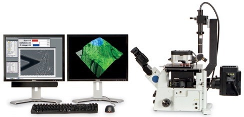
Image Credit: Asylum Research - An Oxford Instruments Company
We chose Asylum’s MFP-3D-BIO AFM because it has the most powerful AFM capabilities of the inverted optical integrated systems. It excels in all aspects, from optical integration to high-resolution imaging and dimensional measurements to force spectroscopy and elasticity measurements of soft tissue matrices. Asylum’s quality and reliability allow us to focus on the science.”
Dennis Discher, Professor, University of Pennsylvania
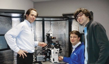
From Left: Dennis Discher, Florian Rehfeldt, Andre Brown. Image Credit: Asylum Research - An Oxford Instruments Company
Performance matters in a bioAFM
Although the term “performance matters” may seem clear, some companies sacrifice performance in their bioAFMs in a variety of ways. Some people have a hole in the center of their AFM skull. While this enables the use of several common optical condensers, it also reduces image resolution and obstructs the measurement of quantitative forces.
In contrast, the MFP-3D-stability BIO enables it to detect tiny characteristics in the samples and quantify picoNewton forces. Since the optical lever mechanism is positioned on a flexure, non-linearities, off-axis movements, and interference fringes are all but eliminated. The specialized condenser optics enable common optical microscopy techniques, such as phase contrast, and offer clear top-down sample viewing.
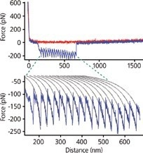
The unfolding of fibril amyloid beta-sheets in algal adhesive reveals a distinct sawtooth pattern. The force curve was fitted to the worm-like chain (WLC) model (dotted lines) and a persistence length of 0.22 nm was calculated. Image Credit: Data from Mostaert et al., J. Biol. Phys. 32(5):393 (2006)
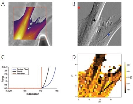
The MFP-3D-BIO includes powerful features for investigating cell mechanics. A) Here, a MRC-five fibroblast cell was located using phase contrast optics and a scan box was drawn to define a region for AFM imaging. The resulting AFM topography data was overlaid on the optical image using thresholding to remove the portion of the image corresponding to the bare petri dish. B) The same optical image was then used to guide the location of three individual force curves. C) The force curves clearly show the stiff petri dish substrate (red), the much softer cell body (black), and a yet softer fiber (blue). D) Finally, a 32 x 32 point force map was measured over the entire area and built-in indentation model analysis was used to calculate the modulus at each point. Image Credit: Asylum Research - An Oxford Instruments Company
Seamless optical and AFM integration
Light up the AFM without compromising performance
The MFP-3D-BIO sets the industry standard for integrating optical and atomic force microscopy in a single integrated tool that is designed specifically for biologists. To guarantee that users gain the maximum benefit and productivity from combining these potent methods, the team of biologists and optical engineers has designed the MFP-3D AFM for use with the top inverted optical microscopes.
Objectives with a high numerical aperture, submersion in water, and TIRF are all supported. The following optical strategies are all supported:
- Phase Contrast
- FRAP
- Brightfield
- FCS
- Confocal Microscopy
- TIRF
- Epifluorescence
- FRET
- Ion Indicators
(e.g. Ca2+ response)
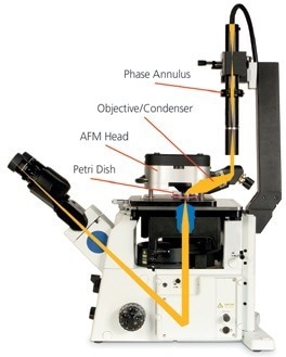
The optical path through the head of the MFP-3D-BIO utilizes a high-quality objective that can be used to view the top of opaque samples and to align the laser onto the tip. The objective can also be used as a condenser for brightfield or phase contrast illumination on transparent samples. High-quality illumination is achieved for observation with the inverted optical microscope objectives without undermining the fidelity of the optical lever or Z-flexure design of the AFM. Image Credit: Asylum Research - An Oxford Instruments Company
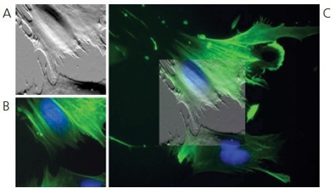
Multiply-labeled fibroblasts imaged in buffer using contact mode AFM (A), and fluorescence microscopy (B). The MFP-3D’s standard overlay feature produced the composite image (C). Our proprietary IR filter blocks the AFM laser, enabling clean, full-spectrum fluorescence imaging—including far-red fluorophores. Image Credit: Asylum Research - An Oxford Instruments Company
The MFP-3D-BIO is a research instrument through and through and designed for the scientist. The optical integration is exceptional, and the flexibility of the platform offers almost endless possibilities.”
Jan Hoh, Professor, Johns Hopkins University
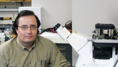
Image Credit: Asylum Research - An Oxford Instruments Company
Illuminate or view with built-in top-down optics
By enabling users to see clear and opaque samples from above while scanning with the AFM, the MFP-3D-BIO expands the capabilities of the inverted optical microscope. Without removing the AFM head or requiring additional equipment, the integrated top-view optics allow in situ laser alignment and tip placement.
The top-view objective lens may serve as a premium condenser for phase-contrast lighting thanks to its innovative design.
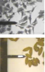
Top-view optical images: (top) HeLa cells on silicon, (bottom) pollen grains. Image Credit: Asylum Research - An Oxford Instruments Company
Real-time optical navigation
It is simple and smooth to maneuver the tip to any feature on the sample, scan that region at the nanoscale with the AFM, or choose certain regions for force curves using top- or bottom-view optical imaging.
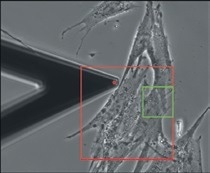
40× phase contrast image of MRC-5 fibroblasts on a Petri dish. With a single click, you can direct the tip (red dot) to any point, or select a new scan area (green box) within the scan range (red box). Image Credit: Asylum Research - An Oxford Instruments Company
Powerful real-time and offline rendering options
It is possible to produce and display combined optical and AFM pictures in real-time and offline. To aid in understanding, optical pictures can be superimposed over AFM data. AFM topography and light microscopy skills are combined in stunning 3D visualizations.
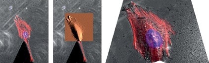
Locate a cell with phase contrast (gray) or fluorescence and examine features such as cytoskeletal structures (red) or the nucleus (purple), then zoom in for high-resolution topography or force measurements with AFM (copper). Overlay optical data on AFM topography for 3D analysis and presentation (right). Image Credit: Asylum Research - An Oxford Instruments Company
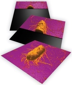
Sequence of real-time 3D renderings of E. coli on glass showing structure and fimbriae, 5 µm scan. Image Credit: Asylum Research - An Oxford Instruments Company
Full capability and ease of use
A bioAFM that satisfies the needs of beginners and experts alike
Even AFM professionals favor keeping operations as straightforward as possible because not all AFM users are experts. Asylum Research creates user-friendly enhancements that benefit both parties in a real way without sacrificing strength or adaptability.
ModeMaster™
- Automatically sets up the program for the chosen mode
- Supports both fundamental and sophisticated imaging methods
- Facilitates rapid and simple start-up
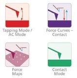
ModeMaster enables one-click configuration for more than thirty different modes. Image Credit: Asylum Research - An Oxford Instruments Company
GetReal™
- The cantilever sensitivity and spring constant are calibrated with one click of the mouse
- There is no chance of tip damage, and the process is quick, easy, and precise
- No need for a firm surface because the tip never makes contact with the sample
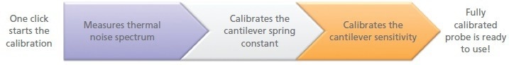
Image Credit: Asylum Research - An Oxford Instruments Company
Automation? Advanced needs? No problem
- By simply dragging “modules” together to create macros with MacroBuilderTM, users may quickly and easily build bespoke functions
- The more basic IGOR Pro scripting language offers much more customization capability
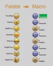
Image Credit: Asylum Research - An Oxford Instruments Company
Our MFP-3D is a highly reliable and key tool in our NanoImaging Core Facility. In addition to ‘traditional’ imaging in air, it has outstanding capabilities for imaging in liquid and performing reliable and accurate force measurements. The simplicity of the software allows us to easily train students, yet the open setup of the instrument makes it flexible and powerful for very high-end projects, including nanolithography and nanomanipulation.”
Yuri Lyubchenko, Professor, University of Nebraska Medical Center
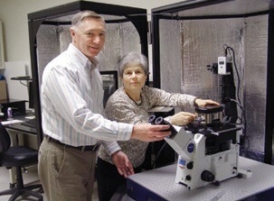
Yuri Lyubchenko with Luda Shlyakhtenko. Image Credit: Asylum Research - An Oxford Instruments Company
Leadership in force spectroscopy and force mechanics
- Thermally-limited force resolution (typical <8 pm deflection noise, guaranteed <20 pm)
- Analysis software assists by recommending the best indentation model from a wide range of pre-built alternatives, such as Hertz/Sneddon, Johnson/Kendall/Roberts (JKR), Derjaguin/Müller/Toporov (DMT), and Oliver-Pharr. Customers may also freely insert their customized models
- Demanding applications, such as cell–cell and cell–substrate adhesion tests, may be accommodated with a large Z range (15 µm standard, 40 µm extended Z option)
- Users can select between closed-loop Z for the most precise velocity control and open-loop force curves with sensored Z for the best possible low noise performance
- The unfolding or rupture dynamics are measured via force clamping for single-molecule and bond-rupture force spectroscopy, allowing for direct comparison to theoretical models
- With the automated fitting of indentation models for the determination of elastic modulus and automated adhesion/rupture force analysis, Force Mapping measures force-distance curves at a grid of locations
Great for biology, but also ready for much more
The MFP-3D-BIO is compatible with all of Asylum’s accessories, modes, and settings because it is built on the potent MFP-3D Origin+ standalone AFM. As a result, it may be prepared to perform its duties effectively in any application, including those that fall beyond the typical scope of bioAFMs, such as those involving materials science, microelectronics, and functional materials. No matter how users choose to conduct their study, it will always support the objectives.
Effective, easy-to-use accessories for biological applications
Petri dish holder
A sample plate that has been specifically created for imaging Petri plates of grown cells and other biological materials.
Peltier dish heater
Petri Dish Holder that allows samples to be heated up to 45 °C.
Closed fluid cell
Sealed compartment with apertures for gas or liquid media at the input and outflow.
BioHeater™
Imaging in a closed fluid cell between room temperature and 80 °C.
Fluid cell lite
An economical fluid cell without ports. Ideal for individual users at multi-user facilities.
CoolerHeater
Uses a Peltier element to heat and cool samples. 24/7 temperature control between –30 °C and +120 °C.
Humidity sensing cell
Measures the humidity within a cell that has been tightly sealed.
MicroFlow cell
Fluid exchange cell with a small volume.
Specifications
High precision 3D motion
All three axes have closed-loop sensors with an X and Y range of 120 µm and a noise level of 0.6 nm.
Z range >15 µm, noise from the Z sensor 0.25 nm Optional Range >40 µm DC height noise for extended Z head with <50 pm
Lowest noise single-molecule or cellular force measurements
<15 pm Cantilever deflection noise (typical 8 pm)
Low coherence source: Superluminescent diode (SLD) for baseline without ripples.
Calibration of the cantilever spring constant using the thermal noise, Sader, or GetReal automated cantilever techniques.
Any channel during a force curve may be recorded or triggered thanks to a flexible interface, including amplitude/phase from AC or Dual ACTM mode, user-supplied input voltages, and photon count rate (with optional Digital Access Module).
Analysis of the elastic modulus and automated adhesion are included in the force mapping.
Optical microscope integration
Includes stage unit for attachment on Olympus IX73, IX81, IX83; Nikon Ti-E/U/S, Ti2-E/A/U; or Zeiss Axio Observer versions of an inverted optical microscope.
All common objective lenses are supported, including high NA TIRF, oil, and water immersion objectives.
For compatibility with far-red fluorophores, an infrared source of 860 nm SLD is used. The SLD beam from fluorescence detectors and cameras is blocked by matched interference filters in the AFM head and optical microscope.
AFM data is overlaid by software with optical pictures in both 2D (varying alpha) and 3D. (topographic rendering).
Region of interest (ROI) selection for imaging and force curves/maps using optical guidance.
For SLD spot alignment and tip placement on opaque materials, top view optics with a 10× / 0.28 NA objective lens are used. For Ph2, Ph1, and Ph0/C/L targets, phase contrast is enabled with an integrated condenser and annuli (purchased separately).
Convergent optical methods for further information on various methods are available.
Superior usability
Integrated modes Contact mode: Dual ACTM, DARTTM PFM; Dual AC Resonance Tracking (DART), Contact mode, Electric force microscopy (EFM), Kelvin probe force microscopy (KPFM), Lateral force mode, Loss tangent imaging, Magnetic force microscopy (MFM), Piezoresponse force microscopy (PFM), Phase imaging, Switching spectroscopy PFM, Fluid imaging, Force modulation, Frequency modulation, Piezoresponse force microscopy (PFM).
Nanolithography/ AC mode tapping; AC mode tapping with Q control; Vector PFM Alternative modes Conductive AFM (CAFM) with EclipseTM mode; AM-FM Viscoelastic Mapping Mode;
Resonant Contact Force modulation, Electrochemical Strain Microscopy (ESM), and Viscoelastic Mapping Mode; Scanning Thermal Microscopy (SThM), High Voltage PFM, Scanning Tunneling Microscopy (STM).
The typical sample format is 75 × 25 mm. A maximum of ×5 mm height by 80 mm in diameter with optional leg extenders, the height may reach 22 mm.
For coverslips (12 or 25 mm diameter or 22 × 22 mm) and Petri dishes, sample adapters are provided (plastic and coverglass-bottom).
Service and Support
One-year comprehensive warranty. Ask about service and support agreements that extend the original warranty and offer additional training and support services.