Materials and Methods
We conducted a retrospective review of 26 patients that had failed total knee replacements treated by knee arthrodesis at a single institution over a 10 year period from January 1981 to December 1991. Average follow-up was 4 years with a range of 2 to 10 years. In 13 patients, an external fixator was used for fixation during the primary arthrodesis procedure. In the other 13 patients, intramedullary nail fixation was used during the primary arthrodesis procedure. Fixation method choice was determined by the operating surgeon. In addition, 6 repeat arthrodeses with intramedullary nail fixation were performed for the treatment of nonunions after external fixation. Union of the arthrodesis site was defined as bony trabecular bridging between the tibia and the femur on plain radiograph.
Seven women and 6 men underwent external fixation arthrodesis for failed total knee replacements. The mean age of this population was 69 years with a range of 51 to 84 years. The underlying diagnoses before the initial total knee replacement was osteoarthritis in 11 patients and rheumatoid arthritis in 2.
Twelve of these patients had condylar type implants and one had a fixed hinge type implant that developed aseptic loosening with severe bone loss. One of the condylar type implant patients also had aseptic loosening associated with painful, limited range of motion and a previous patellectomy. Two of the 13 knees (in 13 patients) treated with external fixation had aseptic failure of their total knee arthroplasty.
The remaining 11 patients in the group undergoing external fixation had septic failure of their total knee arthroplasty. A single causative pathogen was isolated on preoperative aspiration or intraoperative culture in six knees. These inciting organisms included those of the genera Proteus, Pseudomonas, Enterococcus, as well as Streptococcus viridans in one patient each and Staphylococcus aureus in the other two patients. Multiple organisms grew from cultures in the remaining five patients of the 11 who experienced septic failure. These inciting organisms were identified as Staphylococcus epidermidis and Pseudomonas spp in one patient; Staphylococcus aureus and Pseudomonas spp in one patient; Staphylococcus aureus and Peptostreptococcus spp in two patients; and a combination of Staphylococcus aureus, Escherichia coli, Peptostreptococcus spp, and Klebsiella spp in another patient.
The septic cases underwent an average of two surgical debridements (ranging from one to five) prior to the definitive arthrodesis procedure with external fixator placement. At the time of arthrodesis, total knee instrumentation was often used to assist in achieving correct alignment and bone apposition. In addition to the debridements, one patient had a gastrocnemius local muscle flap and four patients had iliac crest bone grafting to supplement bone graft obtained from local sources. Arthrodesis procedures were performed under tourniquet. Seven patients had a uniplanar fixator of the Hoffmann-Vidal type.[6] Another six patients had biplanar fixators in which the Hoffmann-Vidal frame was modified with additional sagittal pins connected to the main frames. These fixators were left in place for a mean of 12 weeks (range, 8 to 14 weeks) followed by long leg casting, until there was evidence of union on radiograph.
In 13 patients, an intramedullary nail was used for fixation of the femur to the tibia. Three men and 10 women comprised this group with a mean age of 55 years (range, 34 to 72 years). The underlying diagnosis prior to the initial arthroplasty was primary bone tumor in two patients, rheumatoid arthritis in one patient, and osteoarthritis in 10 patients. There were two constrained hinge type prostheses in this group and two unicondylar replacements. Nine patients had condylar type total knee replacements. Six patients in this group had aseptic failure of their total knee replacement and in addition to aseptic loosening, four patients had severe bone loss, one patient had limited range of motion with patellectomy, and one patient with a patellar avulsion had a painful, limited, range of motion. Seven patients in this group had septic failure of their total knee replacement and the causative pathogens were identified by aspiration preoperatively or via intraoperative cultures. S epidermidis was the single inciting organism isolated in four patients, and S aureus was the single organism isolated in two patients. A combination of Staphylococcus aureas and Pseudomonas spp was isolated in one additional patient.The technique for arthrodesis of the knee with intramedullary fixation that we used has been described in detail by Vander Griend.[17] In summary, nail size is determined preoperatively by full length anteroposterior and lateral radiographs with a metric radio-opaque ruler placed at the level of the bone. A closed circular, externally fluted Sampsonreg. nail (Kirschner Medical) was used for intramedullary nail fixation in all the cases except one (Fig. 1A,B). This one patient had a 25cm massive intercalary allograft to fill a large bony defect left behind after a failed constrained hinged custom distal femoral tumor endoprosthesis. In this case, a custom ordered proximally and distally interlocked Richardsreg. nail was used (Fig. 2A-C). The allograft used in this case was a fresh frozen femoral shaft. The size of this allograft was selected based on preoperative radiographs. The Sampsonreg. nail has a precurved proximal portion to accommodate the anterior bow of the femur while the distal portion of this nail is straight to accommodate the intramedullary canal of the tibia. As detailed by Vander Griend,[17] the femoral and tibial canals are first reamed 2mm larger than the nominal nail size. The tibia is reamed first because the diameter of the nail is generally determined by the isthmus of the tibial canal. After reaming, the nail is passed retrograde in the femur with the leg flexed and adducted to allow the rod to pass through the trochanter and out through a small incision made in the buttock. When the nail is flush with the distal end of the femur, the tibia is placed against the femur in the position of final alignment, making sure there is good bony apposition and that rotational alignment is correct. Then the nail is driven antegrade into the tibia while axial pressure is held on the patient's foot to prevent distraction of the tibia at the arthrodesis site. We perform most of this procedure using a sterile tourniquet in order to minimize blood loss.

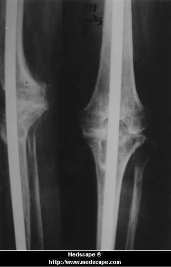
(A). Radiograph of a painful total knee arthroplasty in a 68-year-old woman infected with S aureus and Pseudomonas spp; (B) Radiograph of the same patient six weeks after arthrodesis with a Sampson rod. The patient had three debridement procedures prior to the arthrodesis.

(A). Radiograph of a painful total knee arthroplasty in a 68-year-old woman infected with S aureus and Pseudomonas spp; (B) Radiograph of the same patient six weeks after arthrodesis with a Sampson rod. The patient had three debridement procedures prior to the arthrodesis.

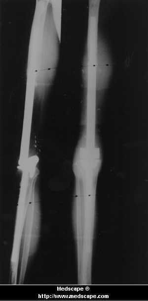
(A) Radiograph of a 34-year-old woman's knee with an S epidermidis-induced infection 10 years after placement of a Guepar hinge tumor prosthesis; (B) Radiograph of the same patient three months after two debridement procedures with placement of a temporary antibiotic PMMA cement spacer and IM rod; (C) Radiograph taken four months after an intercalary allograft arthrodesis using a custom interlocked Richards nail. There was clinical and radiographic union at nine months.

(A) Radiograph of a 34-year-old woman's knee with an S epidermidis-induced infection 10 years after placement of a Guepar hinge tumor prosthesis; (B) Radiograph of the same patient three months after two debridement procedures with placement of a temporary antibiotic PMMA cement spacer and IM rod; (C) Radiograph taken four months after an intercalary allograft arthrodesis using a custom interlocked Richards nail. There was clinical and radiographic union at nine months.

(A) Radiograph of a 34-year-old woman's knee with an S epidermidis-induced infection 10 years after placement of a Guepar hinge tumor prosthesis; (B) Radiograph of the same patient three months after two debridement procedures with placement of a temporary antibiotic PMMA cement spacer and IM rod; (C) Radiograph taken four months after an intercalary allograft arthrodesis using a custom interlocked Richards nail. There was clinical and radiographic union at nine months.
In the seven cases with septic failure, an average of two debridement procedures (range, one to three) were used to debride all the prostheses and bone cement prior to the staged definitive arthrodesis procedure. After debridement and removal of the prosthesis, patients were left with a resection arthroplasty until the intramedullary nail arthrodesis was performed. A PMMA cement-antibiotic spacer mixed with tobramycin and vancomycin was used in all seven patients after the initial implant removal. The time between the final debridement procedure and the intramedullary nail arthrodesis averaged 22.6 weeks (range, 6 to 40 weeks). Prior to proceeding with intramedullary nail arthrodesis, it was required that the patient had negative intraoperative cultures or that aspiration of the resection arthroplasty showed no growth of organisms while the patient was off antibiotics. All seven patients also had sedimentation rates that were less than 20mm/hr prior to the arthrodesis procedure.
In addition to the above procedures, two patients in this group had local gastrocnemius rotation flaps for soft tissue coverage and two patients had intercalary structural allografts for massive bone loss. Three patients with intramedullary nail fixation had iliac crest bone grafting.
Six patients from the external fixation group developed painful nonunions of their attempted arthrodesis. Two of these patients had recurrence of acute sepsis and underwent an additional four debridements each in addition to a local gastrocnemius rotation flap for soft tissue coverage. The inciting organisms were those originally involved in the failed arthroplasty. In one patient, Pseudomonas spp was the causative pathogen and in the other, a combination of Pseudomonas spp and S epidermidis was isolated. The recurrent infections were controlled with debridements and appropriate IV antibiotics and after negative cultures were obtained, these patients underwent repeat arthrodeses using intramedullary nail fixation. Iliac crest bone grafts and Sampson rods were used in all six patients. Two of the patients underwent iliac crest bone graft a second time before union was achieved. These two grafting procedures were performed 13 and 35 months following the intramedullary arthrodesis.
Functional assessment was determined by measuring pain in the affected extremity, walking ability, restriction of activity, the need of external support for ambulation, and patient satisfaction. Patients were classified as having an excellent result when they had no pain, felt that their activities were not restricted, required no external support for walking and were satisfied with their extremity. Patients were considered to have good results if they had occasional modest pain that did not necessitate medication or they needed to use a cane for ambulating distances greater than two or three blocks. A fair result was defined as moderate pain necessitating occasional medication and the need for a cane or walker for any significant ambulation. Patients with poor results had continuous pain requiring frequent medication and were limited to ambulating indoors with the aid of a walker or crutches. Patients who were dissatisfied with having a fusion were considered to have a poor result.
© 1999 Medscape
Cite this: Knee Arthrodesis in the Treatment of Failed Total Knee Replacement - Medscape - May 01, 1999.