Integrated Wearable Ultrasound: Deep Tissue Monitoring in Motion
05-06-2023 | By Robin Mitchell
Recently, researchers from the University of California published their findings on a newly developed device capable of monitoring critical heart functions via ultrasound. What challenges do previous ultrasonic devices face, what did the researchers develop, and how could it be beneficial for medical applications in the future?
What challenges do mainstream ultrasonic devices face?
Ultrasound has proven itself to be immensely popular in the field of medicine for numerous reasons. One such reason is that it allows for internal body images (both 2D and 3D) to be taken without the need for any invasive procedures, thus entirely eliminating the chance of infection and secondary complications. The lack of invasive procedures also means that less skill is required to operate ultrasonic imaging devices, thereby reducing the barrier to entry. This has the impact of reducing their cost to patients while also making them more readily available as nurses can alleviate the pressures on imaging departments.
Another major factor that makes ultrasonic systems ideal for medical imaging is the lack of ionising radiation that other technologies can often introduce (such as CAT and PET scans). This means that patients are not exposed to potentially harmful radiation, and there is no need for specialised shielding or decontamination procedures after each session.
However, for all the advantages that ultrasonic imaging presents, it is not without its flaws. One major flaw that ultrasonic imaging faces is its low resolution compared to other technologies, meaning that it can be challenging to spot small features. At the same time, the limited penetration capabilities of ultrasound waves make it difficult to observe deep tissue, thus restricting imaging to subsurface features.
Another challenge that ultrasonic imaging faces is the need for a cable connected to a large machine used to process data coming from the ultrasonic transducer. This makes portable ultrasonic devices practically impossible using current technology, thereby restricting its use to hospitals with all necessary infrastructure.
To further illustrate the technical challenges, it's important to understand that the current ultrasonic devices face issues related to their size and flexibility. The devices are often rigid and bulky, making them uncomfortable and impractical for long-term wear. Moreover, they require a wired connection to a large machine for data processing, which limits their portability and convenience.
Researchers create wearable ultrasonic device for monitoring heart vitals
Recognising the advantages that ultrasonic systems can provide, researchers from the University of California recently published their findings on a newly developed wearable device that is capable of monitoring key heart functions such as blood pressure and heart rate.
While wearable devices already exist for monitoring heart rate and blood oxygen, none so far can measure vitals such as blood pressure, and this primarily comes down to the difficulty in monitoring pressure without restricting blood flow (typically, blood pressure is monitored by determining the pressure needed to stop blood flow in the arm). At the same time, other wearable ultrasound devices have required wires connected to external devices, thus making them unsuitable for portable use.
However, the researchers were able to develop a small ultrasonic system integrated into a patch that, once placed over the chest, can collect data and transmit these readings to a remote device wirelessly. Once this data is received by a smartphone, AI is used to recover key vitals, thereby allowing for long-term tracking of heart health. Simultaneously, the new device is also capable of sensing as deep as 164mm into the body, making it practical for monitoring organs such as the heart.
As Hongjie Hu, a postdoctoral researcher in the Xu group and study coauthor, explains, "We invented a wearable device that can frequently evaluate the stiffness of human tissue. In particular, we integrated an array of ultrasound elements into a soft elastomer matrix and used wavy serpentine stretchable electrodes to connect these elements, enabling the device to conform to human skin for serial assessment of tissue stiffness." This innovative design allows for a more comfortable and practical user experience.
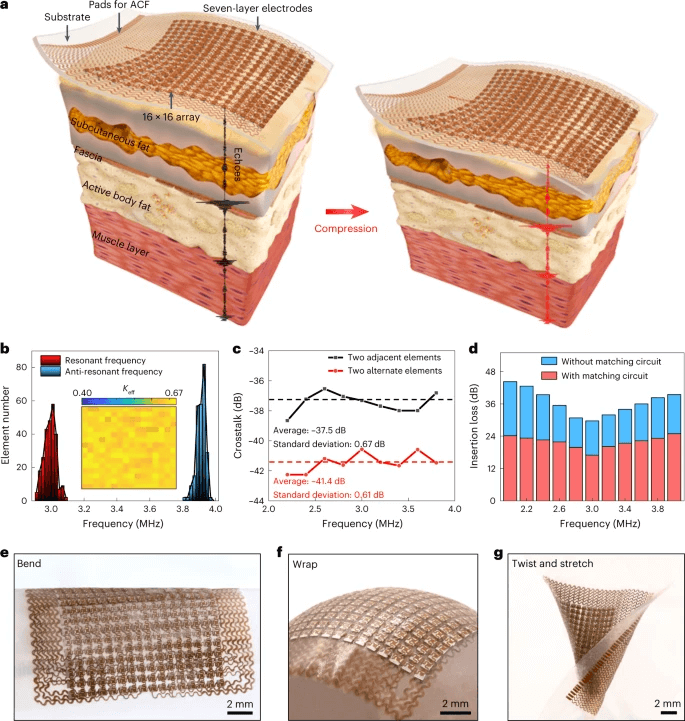
The stretchable ultrasonic array, as depicted in Figure 1, is a sophisticated device designed for application on soft tissue. It comprises a 16-by-16 array of transducer elements (a) interconnected by a seven-layer electrode and encased in a waterproof, biocompatible silicone elastomer. This configuration facilitates individual activation of each element with specific time-delay profiles, enabling the capture of reflected echoes from scattering sources both pre- and post-compression. The signals obtained are then cross-correlated to determine displacement, strain, and modulus fields. The device's resonant frequency, anti-resonant frequency, and effective electromechanical coupling coefficient (Keff) are distributed across the 256 elements (b). The crosstalk between adjacent and alternate elements has been evaluated (c), as well as the insertion loss of the transducers with and without the matching circuit (d). The device demonstrates remarkable mechanical compliance and robustness, maintaining functionality when bent, wrapped on complex-shaped surfaces, or twisted and stretched (e-g).
According to a study published in Nature Biomedical Engineering, the stretchable ultrasonic array developed by the researchers has shown promising results in testing on various artificial phantom models and ex vivo biological specimens. The technology was able to track the recovery progress of muscular injury in a non-invasive serial manner, providing therapeutic guidance.
The device developed by the researchers is a significant advancement in the field of wearable medical technology. It's a stretchable ultrasonic array that can conform to the skin, allowing for the three-dimensional imaging of tissue modulus at depths of up to 4 cm. This is a leap forward compared to the qualitative strain distribution obtained with conventional quasi-static elastography.
The small size of the device, combined with its partial flexibility, makes it ideal for directly attaching to the skin regardless of if a user is stationary or in motion. Thus, this device will also be able to constantly monitor key health markers during critical times, thereby allowing to identify abnormalities as they occur.
The convenience and accessibility of this device cannot be overstated. As Hu puts it, “This allows patients to continuously monitor their health status anytime, anywhere.” This constant monitoring could potentially lead to earlier detection of health issues and more timely interventions.
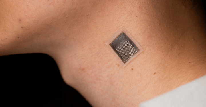
Images showcasing the device's adaptability and durability when twisted and stretched, demonstrating its ability to conform to the lateral shoulder joint and neck for tissue stiffness monitoring. Courtesy of Jacobs School of Engineering, University of California San Diego.
How could such devices be beneficial to future medical applications?
Undeniably, these kinds of devices will soon become commonplace as constant health monitoring allows for long-term trends to be identified. Having such access to data not only helps to catch diseases before they become problematic but will also provide medical AI diagnostic systems with invaluable data. When combined with various other sensors, it is likely that every individual in society will have their own personal GP that never sleeps and always looks out for the tiniest deviations in health.
But these types of sensors are also giving rise to a new industry; the Internet of Medical Things (IoMT). As more and more sensors become connected to the internet, it becomes possible to gather medical data of an individual across the globe. One such possible application where IoMT could provide benefits is food consumption tracking. Whether it is placing an order at a restaurant or weekly grocery shopping, the caloric and nutrition data from consumed food could be pushed to an online cloud service along with all other wearable medical devices, and this could help provide AI-powered GPs to provide better diagnostics and medical advice.
The potential applications of this device are vast. As Gao, another researcher in the group, notes, “Our device shows great potential in close monitoring of high-risk groups, enabling timely interventions at urgent moments.” This could be particularly beneficial for individuals with chronic health conditions or those at high risk of certain diseases.
It's important to note that while the potential benefits of this technology are significant, further research and testing are needed to fully understand its capabilities and limitations. For more detailed information on the research behind this technology, you can refer to the original study published in Nature Biomedical Engineering.
Overall, what the researchers have developed is truly exciting for the field of medical science as well as engineering. It is likely that ultrasonic wearable devices will soon become a commercial reality, but most likely in the form of a chest band that eliminates the need for adhesives.
References:
-
University of California San Diego. (2023). Leading Wearable Ultrasound Lab Creates a Breakthrough in Deep Tissue Monitoring. Retrieved from https://today.ucsd.edu/story/leading-wearable-ultrasound-lab-creates-a-breakthrough-in-deep-tissue-monitoring
-
Hu, H., Ma, Y., Gao, X., Song, D., Li, M., Huang, H., Qian, X., Wu, R., Shi, K., Ding, H., Lin, M., Chen, X., Zhao, W., Qi, B., Zhou, S., Chen, R., Gu, Y., Chen, Y., Lei, Y., Wang, C., Wang, C., Tong, Y., Cui, H., Abdal, A., Zhu, Y., Tian, X., Chen, Z., Lu, C., Yang, X., Mu, J., Lou, Z., Eghtedari, M., Zhou, Q., Oberai, A., Xu, S. (2023). Stretchable ultrasonic arrays for the three-dimensional mapping of the modulus of deep tissue. Nature Biomedical Engineering. Retrieved from https://www.nature.com/articles/s41551-023-01038-w