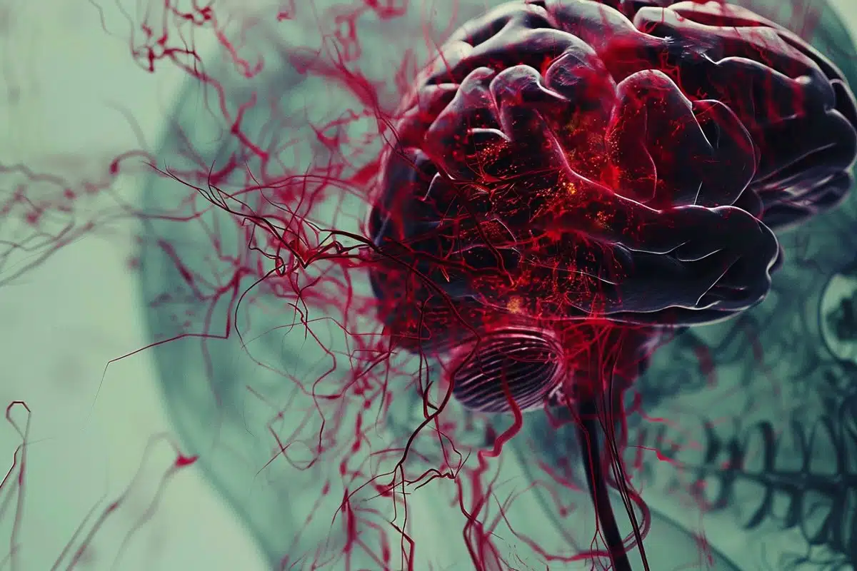
Summary: Microglia, the central nervous system’s immune cells, are crucial in helping the brain recover from anesthesia. By interacting directly with brain circuits and inhibitory synapses, microglia work to counteract sedation effects, offering new insights into managing post-anesthesia delirium and hyperactivity.
This discovery highlights the dynamic role of microglia in maintaining brain health and could lead to innovative treatments for anesthesia-related complications. With advanced imaging technologies, researchers are now able to observe these cells’ active surveillance within the brain, shedding light on their protective functions during states of reduced neural activity.
Key Facts:
- Microglia’s Protective Role: Microglia engage with neurons and inhibitory synapses to mitigate the aftereffects of anesthesia, enhancing neuronal activity for brain awakening.
- Addressing Post-Anesthesia Complications: Understanding microglia’s function could help develop methods to reduce delirium and hyperactivity experienced by patients post-anesthesia.
- Advancements in Brain Cell Imaging: Recent technological advancements have allowed scientists to study microglia movement and interactions in real-time, revealing their importance in brain health and function.
Source: Mayo Clinic
According to a Mayo Clinic study published in Nature Neuroscience, the cells that act as the central nervous system’s first line of defense against harm also play a role in helping the brain awaken from anesthesia. This discovery could help pave the way for innovative methods that address post-anesthesia complications.
When coming out of anesthesia, more than one-third of patients can experience either extreme drowsiness or hyperactivity, a side effect called delirium. Mayo researchers found that special immune cells in the brain called microglia can act to shield neurons from the aftereffects of anesthesia to awaken the brain.

“This is the first time we’ve seen microglia enhance and boost neuronal activity by physically engaging the brain circuits,” says Mayo Clinic neuroscientist Long-Jun Wu, senior author of the study.
The researchers observed microglia wedging between neurons and inhibitory synapses, suppressing neural activity under anesthesia. The microglia appear to be trying to shield the neurons to counteract sedation.
The brain consists of a network of neurons that fire and spur activity throughout the body. Neurons are connected by synapses that receive and transmit signals enabling one to move, think, feel and communicate. In this environment, microglia help keep the brain healthy, stable and functioning. Although microglia were discovered more than 100 years ago, it wasn’t until the last 20 years that they became a serious research focus.
In the beginning, scientists only had fixed slides of microglia to examine, which offered still snapshots of these cells. Initially, it was thought that when neurons were not active and the brain was quiet, microglia were less active. Then technology made it possible to observe and study microglia in greater detail, including how they move.
“Microglia are unique brain cells because they have very dynamic processes. They move and dance around as they survey the brain. We now have powerful images that show their activity and movement,” says Dr. Wu.
Microglia (green) move and “dance” around actively monitoring the brain and interacting with a neuron (red).
For several years, Dr. Wu and his team have been leading research into how microglia and neurons communicate in healthy and unhealthy brains. For example, they have shown that microglia can dampen neuronal hyperactivity during seizures from epilepsy.
In 2019, the researchers discovered that microglia can sense when the brain and its activity is being suppressed, for example, by anesthesia. They found that microglia become more active and vigilant when this occurs.
“We now can see microglia increase their surveillance and patrol the brain’s neural activity like a police officer at night responding to suspicious activity when all else is quiet,” Dr. Wu says.
Patients experiencing delirium or agitation when coming out of anesthesia can also feel hyperactive or experience extreme sluggishness. The researchers believe hyperactivity may result from the microglia intervening too much between the neuron and inhibitory synapses.
“If we can explore the role of microglia in various physiological states, such as sleep, we could apply this knowledge to improve patient care in clinical settings,” says Koichiro Haruwaka, Ph.D., lead author of the study and a Mayo Clinic senior research fellow.
Review the study for a complete list of authors, disclosures and funding.
About this consciousness and neuroscience research news
Author: Emily DeBoom
Source: Mayo Clinic
Contact: Emily DeBoom – Mayo Clinic
Image: The image is credited to Neuroscience News
Original Research: Closed access.
“Microglia enhance post-anesthesia neuronal activity by shielding inhibitory synapses” by Alison Satake et al. Nature Neuroscience
Abstract
Microglia enhance post-anesthesia neuronal activity by shielding inhibitory synapses
Microglia are resident immune cells of the central nervous system and play key roles in brain homeostasis. During anesthesia, microglia increase their dynamic process surveillance and interact more closely with neurons. However, the functional significance of microglial process dynamics and neuronal interaction under anesthesia is largely unknown.
Using in vivo two-photon imaging in mice, we show that microglia enhance neuronal activity after the cessation of isoflurane anesthesia. Hyperactive neuron somata are contacted directly by microglial processes, which specifically colocalize with GABAergic boutons.
Electron-microscopy-based synaptic reconstruction after two-photon imaging reveals that, during anesthesia, microglial processes enter into the synaptic cleft to shield GABAergic inputs. Microglial ablation or loss of microglial β2-adrenergic receptors prevents post-anesthesia neuronal hyperactivity.
Our study demonstrates a previously unappreciated function of microglial process dynamics, which enable microglia to transiently boost post-anesthesia neuronal activity by physically shielding inhibitory inputs.