Bleeding and beating heart models created to help train transplant surgeons

Researchers have created hyper-realistic models of diseased hearts and lungs – which can beat, bleed and breathe like the real organs – to help train surgeons to perform transplants.
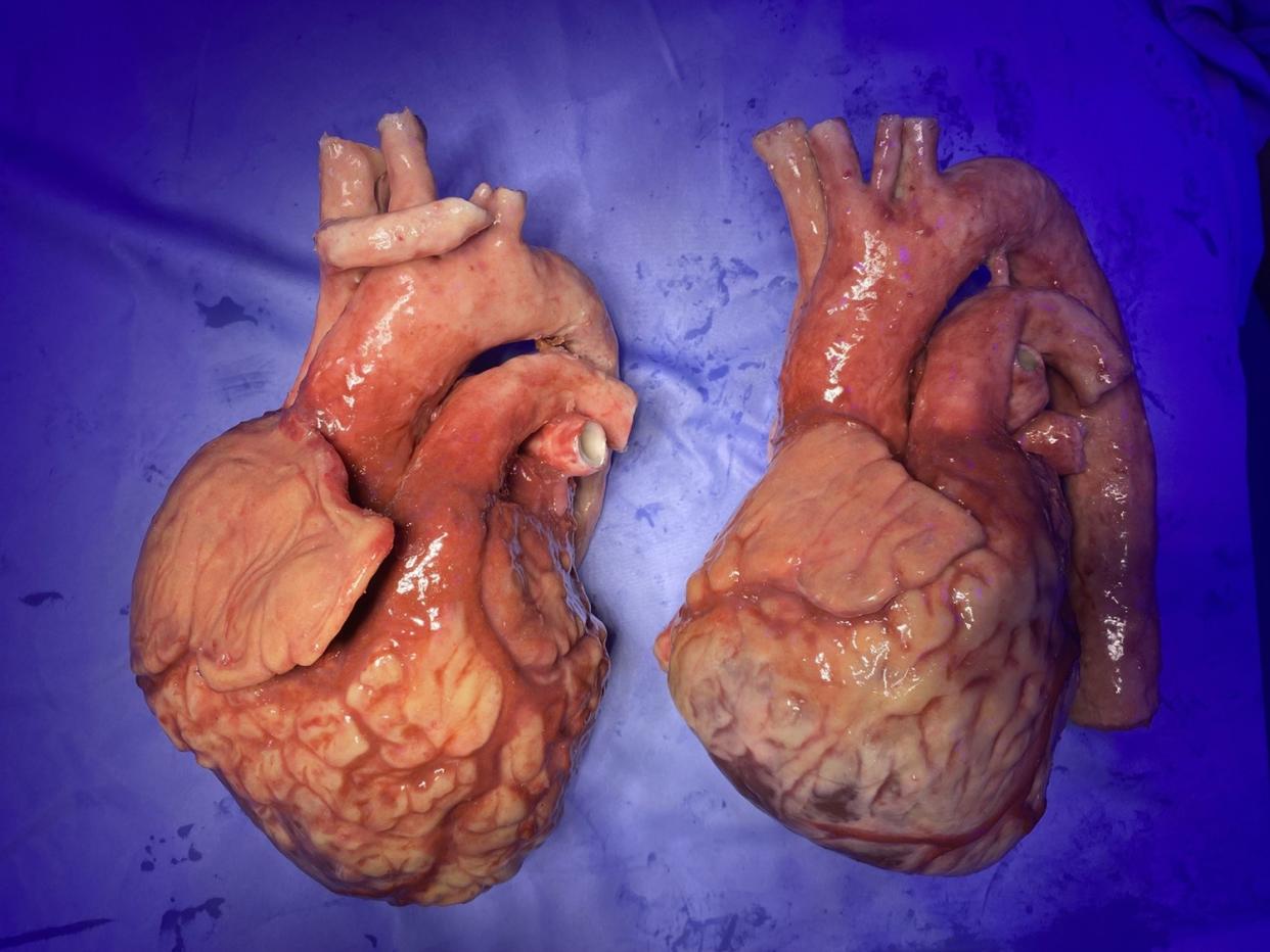
The team, led by senior research fellow Richard Arm at Nottingham Trent University (NTU), said they created the 3D printed fake organs with silicone gels, fabrics and different fibres.
The models, which can be repaired and reused, come with blood vessels that bleed to simulate the real-like experience of clamping them to stem any blood flow.
The organs also have the tactile qualities of human hearts with different levels of tissue hardness, the designers said.
Doctors performing mock surgery can make incisions and remove the diseased organ before suturing a new heart into place with real surgical instruments.
To create the models, the team used heart scans from a patient with heart disease as well as a healthy donor.
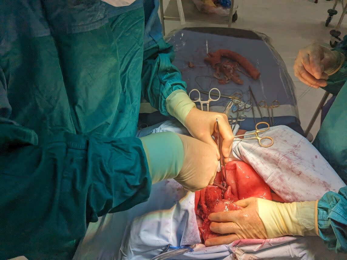
Dr Arm, of NTU’s School of Art & Design, said: “The aim is to give surgeons the opportunity to learn the technical aspects of organ transplant surgery and experience the tactile aspects of removing a failing heart and connecting a different healthy one, identifying and suturing the vessels that keep the donor heart in place.
“This technology can simulate bleeding like a normal heart to provide the actual experience and limited visibility that surgeons must face on the operating table.
“The model is designed to be affordable, reusable and portable, to maximise access to the technology, allowing for increased risk-free training opportunities for transplant surgeons around the country.”
Surgeons will also be able to practice making incisions through the pericardium – a thin sac that surrounds the heart – and the blood vessels which connect the heart to the lungs and the rest of the body.
Dr Arm said earlier versions of the models are being used by the British military and civilian hospitals to improve emergency trauma treatment training.
He said: “While we have made other ‘fake’ organs before, this is our most advanced yet and represents the first example of the next generation in synthetic organs where realistic internal detailing crucially includes the all the important, hidden blood vessels and heart valves.”
“Surgeons currently learn with cadavers and animals because existing models just aren’t realistic enough.
“There’s simply nothing like this in the world right now, so we’re currently working alongside British surgeons and global retailers to trial this next generation of medical models.
“It is really important because heart transplantation is a specialist skill.
“Other examples where our research is making an impact is in maxillofacial prosthetics departments across the UK who use our most advanced skin models to help train plastic surgeons advanced suturing techniques.
“We also produce the world’s most advanced dental models that are used by British dental students to train how to probe gums safely.”
The project, funded by Freeman Heart and Lung Transplant Association (FHLTA), is being presented at the Society for Cardiothoracic Surgery on March 18.
Adele Lambert, chairperson at the FHLTA, said: “The FHLTA are proud to help fund this project as we look to the future of transplantation, we know that this innovative research will help to improve surgeons’ techniques for organ transplantation.”
