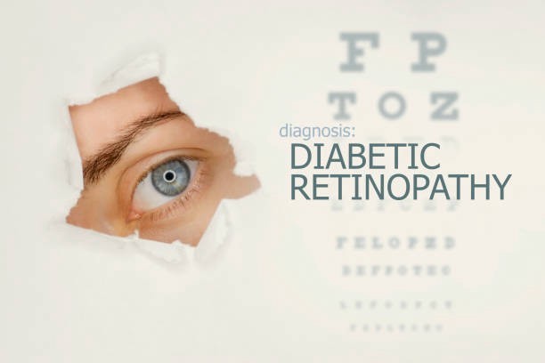
Artificial Intelligence Detects Diabetic Retinopathy in Real–Time
Thanks to advanced algorithms, we can predict and detect various diseases. Thanks to Artificial Intelligence (AI) and Big Data, significant advances have been made in the eye and vision care. However, millions of people suffer from diabetic retinopathy, the leading cause of blindness among working–age adults. In many rural areas of the world, it is hoped that this disease, for which medical screening is complex, will be detected and prevented with new technologies.

The U.S. Food and Drug Administration (FDA) has approved the first medical device that can be used by eye care professionals using Artificial Intelligence (AI) software.
Google has made the system usable in real life by placing an algorithm in the Artificial Intelligence (AI) system that it has been working on for four years to diagnose Diabetic Retinopathy (DR), which damages eyesight. According to ophthalmologists, if the use of this system becomes widespread, a significant decrease in the number of visually impaired people can be observed around the world.
Diabetic Retinopathy, which causes more than 415 million people worldwide to be permanently wholly or partially blind, is an eye disease that causes damage to the retina due to high blood sugar.
For nearly four years, Alphabet’s life sciences company Verily and Google’s Artificial Intelligence research unit have been working on a system to diagnose Diabetic Retinopathy. Google developed an algorithm for this system to be used in real life, and it is currently employed at Aravind Eye Hospital in Mumbai.
Aravind Eye Hospital has been working on the automatic diagnosis of Diabetic Retinopathy since 2003. The hospital’s chief physician, Dr. Kim, said that Google’s Artificial Intelligence (AI) system is far ahead of the system they use in diagnostic speed and accuracy. For example, when diagnosing Diabetic Retionapathy, the Artificial Intelligence (AI) system uses a database of 128,000 images of pupils evaluated by ophthalmologists. The design provided 97.5% accuracy in diagnosing the disease as a result of two separate tests with approximately 12 thousand images.

It is Expected to be Prominent in Different Diseases
Information about this is increasing day by day, and it is starting to be used in the field in various eye diseases. For example, the OCT device used in diagnosing glaucoma (eye pressure disease) detects the nerve fiber thickness of the optic nerve. Thinning of the fibers is also essential in the follow–up of glaucoma. It is thought that diagnosis by Artificial Intelligence (AI) with images obtained from OCT will be possible. In addition to making nerve fiber thickness analysis with OCT, it can also be determined whether there is any problem in the yellow spot in the eye. One is age–related macular degeneration, namely “yellow spot disease.” There are structures defined as “drusen” in the yellow area in this disease. The dimensions of these structures can be followed very quickly with OCT. Although the FDA does not currently approve it, it is thought to be put into clinical use shortly. Apart from this, there is also a picture of edema due to diabetes in the yellow spot. Some measurements are made regarding the laser intervention, in which case the payment is made. Artificial Intelligence (AI) about these measurements will be of great benefit to us. There may be edema in the yellow spot due to some vascular occlusions in the retina. Artificial Intelligence (AI) will also help us decide the amount of these and whether they require treatment. If we come from the retina towards the front of the eye, it is thought to be prominent, especially in detecting congenital cataracts.
Artificial Intelligence (AI) has been on the rise in the last ten years and has come a long way in medicine, radiology, and dermatology. The rapid development in the field of eye diseases continues.
Acıbadem University Atakent Hospital Ophthalmology Specialist Assoc. Dr. Ali Rıza Cenk Çelebi stated that Artificial Intelligence (AI) had come a long way in the diagnosis of anterior and posterior segment diseases of the eye, but there are more to be overcome in terms of treatment, “Artificial Intelligence applications; It can make the diagnosis of Diabetic Retinopathy with fundus images with an accuracy similar to that of an ophthalmologist, and in a faster time than that. Another of the ophthalmological studies on this subject is on glaucoma. Accurate detection, especially in the early stages of this disease, can also prevent the declining quality of life by preventing irreversible vision loss that may develop due to the disease. Patients cannot realize the situation, especially in this disorder, which goes with the loss of the peripheral visual field. On the other hand, Artificial Intelligence can improve the patient’s quality of life by preventing damage from the beginning with early diagnosis. It also gives high accuracy results in the Diagnosis of Retinopathy of prematurity and keratoconus.”
The IDX–DR device, developed by IDx LLC, is the first device authorized for marketing, typically involved in eye care processes and who make a screening decision without the need for a doctor to interpret the image or the results. According to researchers, early detection of Retinopathy is an integral part of managing the care of millions of people with diabetes. Still, many patients with diabetes are not adequately screened for Diabetic Retinopathy because about 50 percent of them do not see their ophthalmologist annually; The IDX–DR device, retina captured with the Topcon NW400 retinal camera. Instead, a doctor uploads digital images of a patient’s retinas to a cloud server with IDX–DR software. Suppose the digital images are of sufficient quality. In that case, the software provides the doctor with one of two results: (1) “more than mild Diabetic Retinopathy detected: Consult an eye care professional for more than mild Diabetic Retinopathy” or (2) “more than mild Diabetic Retinopathy detected; 12 months Repeat viewing in.” In the survey, IDX–DR correctly identified the presence of mild Diabetic Retinopathy more than 87.4% of the time and correctly identified patients who had no more than mild Diabetic Retinopathy less than 89.5%.
Diabetic Retinopathy Detection
- Blurred Vision
- Radiation Retinopathy
- Permanent Vision Loss
- Moving Spots in the Eye
- Retinal Vessel Occlusion
- Proliferative Retinopathy
- Severe Nonproliferative Retinopathy
- Previously Diagnosed Macular Edema
The IDX–DR had a breakthrough device designation and was reviewed through the FDA’s de novo premarket review route, a regulatory pathway for certain low to medium risk devices with a new quiet regulatory environment and no previously legally marketed device. Experts think this new validation channel will pave the way for more innovative devices in this field.