Background
Thrombophlebitis involves the formation of a blood clot in the presence of venous inflammation or injury. Many innate conditions may predispose patients to thrombophlebitis by means of a variety of hypercoagulopathy syndromes. [1] Traumatic events can also initiate a thrombophlebitic reaction. In addition, the persistence of significant reflux into a vein that has been treated with a sclerosing agent can lead to phlebitis. More commonly, phlebitis occurs if perforator veins in the region of sclerotherapy are not diagnosed and treated.
Pathophysiology
Hypercoagulable states
A number of primary and secondary hypercoagulable states can be assessed by obtaining an appropriate patient history and review of systems. Prior to 1993, only 3 inherited hypercoagulable factors had been recognized: antithrombin III, protein C, and protein S. Currently, 60-70% of patients with thrombosis can be identified as having a specific inherited thrombophilia. [2] Inherited hypercoagulable states are divided by experts into 5 main categories: (1) qualitative or quantitative defects of coagulation factor inhibitors, (2) increased level or function of coagulation factors, (3) hyperhomocysteinemia, (4) defects of the fibrinolytic system, and (5) altered platelet function.
The specific inherited thrombophilias are listed below. [3] The majority of these inherited diseases have identified gene mutations, some of which are used in diagnosis. Protein C deficiency alone has more than 160 genetic mutations associated with disease-causing states. [4]
Inherited thrombophilia classifications are described below. [3]
Qualitative/quantitative defects of coagulation factor inhibitors are as follows:
-
Antithrombin deficiency
-
Protein C deficiency
-
Protein S deficiency
-
Heparin cofactor II deficiency
-
Tissue factor pathway inhibitor deficiency
-
Thrombomodulin deficiency
Increased levels/function of coagulation factors are as follows:
-
Activated protein C resistance and factor V Leiden
-
Prothrombin gene mutation (G20210A)
-
Dysfibrinogenemia and hyperfibrinogenemia
-
Elevated levels of clotting factors VII, VIII, IX, XI, and XII
Hyperhomocysteinemia is another class.
Defects of the fibrinolytic system are as follows:
-
Plasminogen
-
Tissue plasminogen activator
-
Thrombin-activatable fibrinolysis inhibitor
-
Factor XIII
-
Lipoprotein (a)
Altered platelet function conditions are as follows:
-
Platelet glycoprotein GPIb-IX
-
GPIa-IIa
-
GPIIb-IIIa
In the general population, the prevalence of an inherited thrombotic syndrome is presently estimated to be 1 individual in 2500-5000 population; the prevalence increases to 4% in patients with a past history of thrombosis. [5] A past history of deep venous thrombosis (DVT) increases the likelihood of new postoperative venous thrombosis from 26% to 68%, whereas a past history of both DVT and pulmonary embolism (PE) is predictive of a near 100% rate of thrombosis. [6] Further detail regarding hypercoagulable conditions is beyond the scope of this article. The most common conditions are discussed below. For additional information, the reader is referred to multiple review articles on hypercoagulable conditions. [3, 5, 7, 8]
Resistance to activated protein C (APC) is the most common genetic risk factor associated with venous thrombosis. Most cases are due to a point mutation in the factor V gene (factor V Leiden FVL]), which subsequently prevents the cleavage and disruption of activated factor V by APC and thus promotes ongoing clot development. In approximately 3-8% of white adults, this mutation is heterozygous, conferring a 5-fold increased lifetime risk of venous thrombosis compared with the general population. [9] Double heterozygosity with FVL and protein C, protein S, or antithrombin deficiency is reported, and affected individuals have an increased risk of thrombosis. Women with FVL heterozygosity who are also taking oral contraceptives have a 35-fold increase in the risk of thrombosis. Homozygotes of FVL have an 80-fold increased risk for venous thromboembolism. [10]
Inherited factor deficiency
Although endothelial damage is speculated to be necessary for symptomatic thrombosis to occur, venous thrombosis may be associated with a deficiency in 1 of several anticoagulant factors. [11] In otherwise healthy patients younger than 45 years who are referred for evaluation of venous thrombosis, the prevalence of antithrombin III, protein C, and protein S deficiency is approximately 5% for each. [12, 13]
Antithrombin (antithrombin III) deficiency occurs in 1 person per 2000-5000 people in the general population and is the most prothrombotic of all inherited thrombophilias. [14, 15, 16] Acquired antithrombin deficiency can occur with liver disease and as a result of oral contraceptive use. Antithrombin combines with coagulation factors, blocking biologic activity and inhibiting thrombosis.
Protein C and protein S, 2 vitamin K–dependent proteins, are other important anticoagulant factors. Protein S is a cofactor for the effect of APC on factors Va and VIIIa. In the United States, the prevalence of heterozygous protein C deficiency is estimated to be 1 case in 60-300 healthy adults. [17] Greater than 95% of the patients are asymptomatic. However, a significant deficiency in either protein can predispose an individual to DVT. In fact, 75% of patients with homozygosity for protein S deficiency have venous thrombosis before age 35 years. [18]
Although factor deficiency can cause venous thrombosis, a genetic alteration in factor V, which results in APC resistance, is at least 10 times more common than other alterations. This genetic alteration is found in approximately one third of patients referred for an evaluation of DVT. [19, 20, 21] Precipitating factors for thrombosis, such as pregnancy and the use of oral contraceptives, are present in 60% of these patients. APC resistance is discussed at the beginning of the Pathophysiology section under Hypercoagulable states.
Defects in the fibrinolytic system, specifically plasminogen, occur in as much as 10% of the healthy population. [22] When the defects occur alone, the risk of thrombosis is small. Under certain circumstances, abnormal plasminogen levels may also predispose an individual to thrombosis.
Antiphospholipid antibodies are a cause of both venous and arterial thrombosis, as well as recurrent spontaneous abortion. [4] They may manifest in a primary thrombophilic disorder, or they may be related secondarily to autoimmune disorders. Lupuslike anticoagulants are present in 16-33% of patients with lupus erythematosus, as well as in many patients with a variety of autoimmune disorders. [23, 24, 25] Thrombosis may occur in 30-50% of patients with circulating lupuslike anticoagulants. [25, 26, 27]
Oral contraceptive use and estrogen replacement therapy
The mechanism for thromboembolic disease in women who use oral contraceptives is multifactorial. Both estrogens and progestogens are implicated in promoting thrombosis, even with low-dose therapy. [28, 29, 30, 31] All study results indicate that the increased risk occurs predominately during the period of use and perhaps for a week or so after discontinuation. [32, 33] However, the total correction of potentially hemostatic changes that occur during oral contraceptive therapy requires 4 weeks of abstinence. [34]
The highest rate of thromboembolism occurs with the use of large doses of estrogen [28, 29, 30, 32, 35] some studies show an 11-fold increase in thromboembolism. [32, 36] Nevertheless, the risk of postoperative PE still appears to be increased in women who use oral contraceptive agents, even with minimal amounts of estrogen. [37]
The incidence of DVT associated with oral contraceptive use varies depending on the type and concentration of estrogen. The potency among native estrogens, estrone and estradiol, ethinyl estradiol, and estrogens in oral contraceptive agents differs by at least 200-fold. [38] In patients who receive hormone replacement therapy with 0.625 mg of conjugated equine estrogens and 2.5 mg of medroxyprogesterone, the risk of DVT is 2-3.6 times higher than that of nonusers. [39]
Oral contraceptives are responsible for approximately 1 case of superficial venous thrombosis (SVT) or DVT per 500 women users per year. [40] This incidence of symptomatic thrombosis may be a low estimate of true hypercoagulability; a plasma fibrinogen chromatographic study demonstrated a 27% incidence of silent thrombotic lesions in 154 new users of either mestranol at 100 mg or ethinyl estradiol at 50 mg. [41]
As a group, people who take oral contraceptives have numerous alterations in their coagulation system that promote a hypercoagulable state. These alterations include hyperaggregable platelets, decreased endothelial fibrinolysis, [42] decreased negative surface charge on vessel walls and blood cells, [43] elevated levels of procoagulants, reduced RBC filterability, [44] increased blood viscosity secondary to elevated RBC volume, [45] and decreased levels of antithrombin. [46, 47, 48] An alteration in any of these factors, alone or in combination, may predominate in women who are taking oral contraceptives. The extent of the derangement in the hemostatic system determines whether thrombosis occurs.
The most important factors that prevent clot propagation are antithrombin and vascular stores of tissue plasminogen activator (t-PA). [46, 49, 50, 51] Antithrombin levels are 20% lower in some women who are taking oral contraceptive agents [49] or estrogen replacement medications. [52] In women who use oral contraceptive agents and have thromboembolic events, releasable t-PA is decreased 25-fold in 90% [46, 49, 50] and the venous walls in 51.6% have an abnormally low plasminogen activator content. [51] Therefore, a certain subgroup of women who are taking birth control pills may have a particular risk for thromboembolic disease.
In addition, the distensibility of the peripheral veins may increase with the use of systemic estrogens and progestins. [53] This increased distensibility may promote valvular dysfunction and a relative stasis in blood flow, both of which enhance the hypercoagulable state.
A therapeutic alternative that should be considered for women in whom estrogen replacement cannot be discontinued is transdermal 17-beta-estradiol. The direct delivery of estrogen into the peripheral circulation eliminates the first-pass effect of liver metabolism. This delivery method decreases hepatic estrogen levels, with subsequent minimization of the estrogen-induced alteration of coagulation proteins. Thus, the use of transdermal estrogen is recommended for patients with an increased risk of thromboembolism because alterations in blood clotting factors have not been demonstrated during such treatment. [54]
Tamoxifen use
Unusual and poorly understood complications of tamoxifen use are thrombophlebitis and DVT. These complications occur in as many as 1% of treated patients. [55, 56] Results from the evaluation of various coagulation parameters and factors, including the sex hormone–binding globulin level, antithrombin activity, fibrinogen level, platelet count, protein C level, and fibrinopeptide A level, are all normal. [56, 57, 58, 59, 60] By contrast, one small case series of women experiencing venous thrombosis found APC resistance attributable to factor V Leiden heterozygous mutations in all 3 patients. [61]
Pregnancy
During pregnancy, an increase in most procoagulant factors and a reduction in fibrinolytic activity occur. Plasma fibrinogen levels gradually increase after the third month of pregnancy, to double those of the nonpregnant state. In the second half of pregnancy, levels of factors VII, VIII, IX, and X also increase. [62] Decreased fibrinolytic activity is probably related to a decrease in the level of circulating plasminogen activator. [63] In addition, a 68% reduction in protein S levels is measured during pregnancy and in the postpartum period. [64] Protein S levels do not return to the reference range until 12 weeks after delivery. These changes are necessary to prevent hemorrhage during placental separation.
The hypercoagulable condition of the immediate antepartum period is responsible, in large part, for the development of superficial thrombophlebitis and DVT in 0.15% and 0.04% of this patient population, respectively. [65] Even more important is the immediate postpartum period, during which the incidences of superficial thrombophlebitis and DVT increase to 1.18% and 0.15%, respectively. A Dutch study of pregnant women with age-matched controls found a 5-fold increased risk of venous thrombosis during pregnancy. This increased to 60-fold during the first 3 months after delivery. [66] Fifty-percent of DVT cases develop by the second day after delivery, and 84% of DVTs in pregnancy occur in the left leg. [67]
Because normalization of most coagulation factors generally occurs by postpartum day 3, [68] additional factors are suspected in the 21% of patients in whom a DVT subsequently develops 2-3 weeks after delivery. Maternal age may also be linked to venous thrombosis, although study results are conflicting; one of the studies found the rate is approximately 1 case per 1000 women younger than 25 years, changing to 1 case per 1200 women older than 35 years. [35]
Two thirds of patients in whom postpartum DVT develops have varicose veins. Thus, in addition to the potential adverse effects on the fetus, sclerotherapy should be avoided near term until coagulability returns to normal 6 weeks after delivery.
Travel-related venous thrombosis
Although the relationship between air travel and DVT was first recognized in 1954, [69] PE was noted to occur in Londoners confined to air raid shelters during World War II. In 1993, Lord and McGrath reported findings of 45 patients in whom venous thrombosis was related to travel (37 by air and 8 by road or rail). [70] Stationary travel for more than 4 hours increases the risk of venous thromboembolism 2-fold, even several weeks beyond the time of travel. [71] Clinical risk factors included previous thromboembolism (31%) and varicose veins (20%).
Lord reported that in 122 additional patients, thromboembolism was associated with prolonged travel. [72, 73] Hypercoagulable factors were isolated in 72% of patients who were tested. The most common factor was protein C resistance, which was found in 47% of patients.
At least one clinical or laboratory risk factor was present prior to travel in greater than 80% of patients who developed DVT after long-haul flights (>8 h), and SVT was diagnosed in 12% of this study group. [74] In most cases, the risk factors could be identified by medical history, without any laboratory testing. The most common risk factors were estrogen use, history of thrombosis, and the presence of factor V Leiden.
Malignancy and illness
Hypercoagulability occurs in association with a number of malignancies, with the classic example being Trousseau syndrome—a thrombotic event occurring prior to an occult malignancy, usually a mucin-producing visceral carcinoma. The pathophysiology of malignancy-related thrombosis is poorly understood, but tissue factor, tumor-associated cysteine proteinase, circulating mucin molecules, and tumor hypoxemia have all been implicated as causative factors. [75] Symptoms suggestive of malignancy should be investigated in individuals without other known risk factors for thrombosis.
Medically ill patients have a 10% chance of developing a DVT, while hospital-acquired DVTs and PEs occur in 10-33% of all hospitalized patients. Thrombophlebitis in this patient population is promoted by a combination of hypercoagulability and venous stasis. [76] Surgery, trauma, and immobilization also predispose to venous thromboembolism formation. Surgery without anticoagulation is associated with an incidence rate of DVT from 15-64%, while as many as 58% of patients entering trauma units have a DVT. [77, 78]
Other factors
Other disease states are associated with venous thromboembolism. Paroxysmal nocturnal hemoglobinuria, nephritic syndrome, and inflammatory bowel disease all are associated with increased risks of thromboembolism. [4] Alteration of the activity of matrix metalloproteinases influences mechanical properties of the vein wall. [79] The rate of peripheral venous thrombophlebitis following intravenous cannulation varies from 10-90%. [80] Newer catheter materials may be less thrombogenic. [81] Thrombophlebitis may also be a complication of medications that interfere with the coagulation pathway, anticoagulant treatment, [82] or infections. [83] Venous function has been suggested to be influenced by genetic factors. [84]
Mondor disease involves thrombophlebitis of the superficial veins of the breast and anterior chest wall. It has been associated with breast or axillary surgery, malignancy, and intense thoracoabdominal exercise training. [85, 86]
Etiology
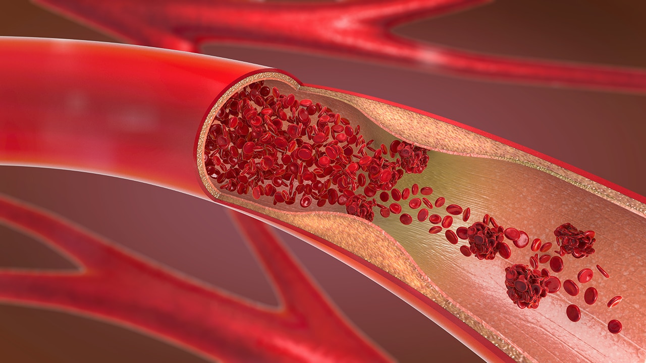
See Pathophysiology.
Trauma to a varicose vein or healthy vein is common.
Predisposing factors include any event that can reduce venous flow; examples include prolonged sitting or immobilization and dehydration (eg, as on a long airline flight), long surgery, or prolonged bed rest.
Genetic thrombophilia or underlying malignancy can lead to a hypercoagulable state.
Internal trauma to a vein due to an indwelling catheter or even a difficult phlebotomy procedure can also cause venous injury and inflammation.
Viral diseases, including coronavirus disease 2019 (COVID-19) and Chikungunya virus infection, have been associated with superficial and deep thrombophlebitis. [87, 88]
Epidemiology
Frequency
The approximate annual incidence of venous thromboembolism in Western society is 1 case per 1000 individuals. [89] The annual incidence of symptomatic venous thromboembolism is decreased compared with asymptomatic, at approximately 0.5 to 1.6 per 1000 individuals. [90] Exact frequency data for the general population are difficult to find. The frequency is influenced by the subgroups of patients studied. Patients with a prior superficial venous thrombosis are at increased risk for deep vein thrombosis. [91] See Pathophysiology.
Race
No racial predilection is recognized.
Sex
Women have a slight predilection over men because of systemic estrogen use.
Age
Age may be a predisposing factor in SVT, DVT, or both. The average age of a European venous thromboembolism registry of more than 15,000 patients was 66.3 ±16.9 years. [92] Reportedly, elderly patients have an increased risk of DVT. [93, 94, 95] The major cause of this increased risk may be the relative pooling of blood in the soleal venous sinuses, which occurs as a result of decreased calf muscle pump infusion. [96]
Prognosis
SVT and DVT both have an excellent prognosis if treated promptly. Proper treatment should result in rapid resolution.
After resolution of the acute problem, the following treatment options for the underlying varicose veins should be considered: ambulatory phlebectomy, ligation and stripping, endovenous radiofrequency ablation, and endovenous laser ablation. [97, 98, 99]
DVT causes edema (79.8%), pain (74.6%), and erythema (26.1%), according to a large Italian registry of patients. [90] It may also be associated with the development of life-threatening PE, if untreated. Similarly, superficial thrombophlebitis is not a complication that should be taken lightly. If untreated, the inflammation and clot may spread through the perforating veins to the deep venous system. This extension may lead to valvular damage and possible pulmonary embolic events. [100, 101, 102, 103, 104] Propagation of SVT to DVT may occur in up to 15% of patients. [105] Alarmingly, 10% of SVT either recurs, extends, or progresses to DVT despite treatment. [106] SVT in the presence of an acquired thrombotic risk factor increases the risk of VT by 10- to 100-fold. [107] Superficial thrombophlebitis is associated with an elevated risk of recurrence. [108]
Coincidental DVT with SVT is reportedly more common in patients without varicose veins than in those with varicose veins (60% vs 20%). Thus, other innate factors place patients with SVT at additional risk for DVT.
In a study of 145 patients, superficial thrombophlebitis in 23% of the affected limbs had proximal extension into the saphenofemoral junction (SFJ). [109] PE was found in 7 (33.3%) of 21 patients with thrombophlebitis of the greater saphenous vein (GSV) above the knee. [110] Seventeen of the 21 patients had varicose veins. In this study, clinical symptoms suggestive of PE were present in only 1 of 7 patients. The occurrence of DVT in patients with below-the-knee SVT was 25 (32%) in a study of 78 patients. [111]
A European registry of 4405 patients with acute venous thromboembolism had a 3.1% rate of adverse events in the 3 months following the initial insult. These adverse events included symptomatic PE (0.3%), recurrent DVT (0.4%), major bleeding (0.8%), and death (1.5%). [112]
Patient Education
Patients should be educated regarding the risk factors for future thrombotic events. The risks and benefits of anticoagulation therapy should also be explained.
For patient education resources, visit the Lung Disease and Respiratory Health Center. Additionally, see the patient education articles Varicose Veins, Blood Clot in the Legs, Phlebitis, and Pulmonary Embolism.
The Journal of the American Medical Association’s patient education information [113] is referenced in the Bibliography.
-
Superficial thrombophlebitis. Courtesy of DermNet New Zealand (http://www.dermnetnz.org/assets/Uploads/vascular/thrombophlebitis.jpg).
-
Deep venous thrombosis.