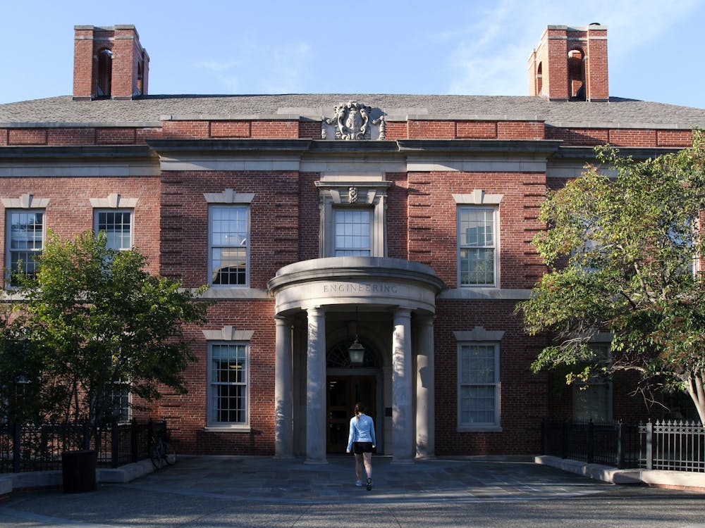
Tucked away in the Engineering Quad, students and scientists watch as syringes pump out a steady stream of translucent beads, no more than 90 microns in diameter. These beads are made of hyaluronic acid, and will soon be manipulated to form a hydrogel, or a synthetic microenvironment for human tissue growth.
This is the lab of Professor of Biomedical Engineering Tatiana Segura, who has spent nearly a decade focusing on repairing tissue in one of the most complex human organs: the brain.
Segura’s work focuses on designing hydrogels to mimic the extracellular matrix in the brain. This matrix is responsible for providing cells nutrients, enzymes and other factors that are critical for growth. When the brain suffers an injury, such as a stroke, the matrix is destroyed alongside the cells, eliminating the possibility of natural regrowth.
“There is this idea that you can implant something [manmade] to take on the function of this provisional matrix, and see if you can keep the repair process going for longer,” Segura said. “But what are the key features of this matrix? This is what we’re struggling with.”
The actual creation of a hydrogel takes barely more than an hour. Segura’s job, however, is to chemically modify each gel in order to suit the needs of patients. Segura and her lab have gone through years of constant modifications in an attempt to balance the promotion of positive growth with the inhibition of worsening symptoms.
In a recent study, the lab found that brain tissue of mice that had suffered stroke injuries regenerated after injecting one of their engineered hydrogels into their brains. However, Segura is hesitant to draw definitive conclusions about the correlation, especially when it comes to applying these results to humans.
Treating humans, she noted, is an entirely different story when it comes to the injection of gels for brain tissue regeneration.
“I think about transport and mechanics as just being a huge problem,” she said. “When you inject a few microliters of gel compared to a few milliliters of gel, the mechanics of that will be different. The diffusion of molecules and nutrients will be a completely different time scale. But that is the goal.”
By collaborating with various other researchers and clinicians, Segura hopes to be able to create therapies for people who have stroke-related disabilities. Currently, no therapies for these types of disabilities exist.
“I think the clues are there,” she said. “We just need to figure out what [genes] need to be activated.”
Segura is not the only Duke researcher utilizing hydrogels to solve the problem of muscle regeneration. Professor of biomedical engineering Nenad Bursac has found that hydrogels are useful for creating entire tissues outside of the body in addition to creating an extracellular matrix that can be injected into the body.
“We remake tissues in the gel, and then we surgically implant the whole tissue inside the body,” Bursac said. “So when we have a defective injury, we replace that lost tissue with engineered muscle that we make.”
His work focuses on the regeneration of skeletal muscular tissue in an in vitro microenvironment. According to Bursac, the goal of his research is to engineer these hydrogels to form environments that will foster muscle growth in patients who suffer from a variety of muscle diseases, including muscle inflammation and genetic diseases like muscular dystrophy.
Like Segura, Bursac works on finding the right combination of enzymes, growth factors and nutrients that will create the right type of nourishing environment for atrophied muscle cells. He has been working on this project for 12 years and has gone through years of optimizations in order to find the key ratios for each of these factors.
The primary struggle he faces is mimicking every component found in the actual human body, such as the nerves that run through each muscle.
“We don’t have every component of a muscle built into these engineered tissues,” Bursac said. “We try to mimic [innervation] through electrical stimulation, but we don’t have neuronal activity or other things that neurons secrete.”
He also echoed Segura’s concern about the sizeable scale difference between mice or rats and humans. Using microgels, Bursac has been able to engineer tissues with the potential to be implanted into cavities muscle disease leaves behind.
But these tissues are often too small to repair the whole injury. Instead, he focuses on developing microtissue that can be used to test novel therapies.
“[These gels] are mostly used as test beds,” he said. “The problem with repairing muscles is that [the injury] would have to be localized to one area, but many diseases affect muscles everywhere. For these systemic injuries, this is used as a test bed to understand the drugs or gene therapies that would potentially help.”
Bursac points to the potential of using engineered tissue environments to reduce animal testing by pharmacological companies in favor of using tissue samples to test products and develop personalized drug therapies based on an individual’s muscle cells. This research could then be applied to new techniques, such as 3D printing.
“For example, 3D printing may allow us to generate large muscle tissues that are viable,” Bursac said. “But there is still a lot of work to be done.”
Get The Chronicle straight to your inbox
Signup for our weekly newsletter. Cancel at any time.

