May 5 2020
Ever since the initial description of a cell in Micrographia made by Robert Hooke, 350 years ago, microscopy has played a significant role in interpreting the rules of life.
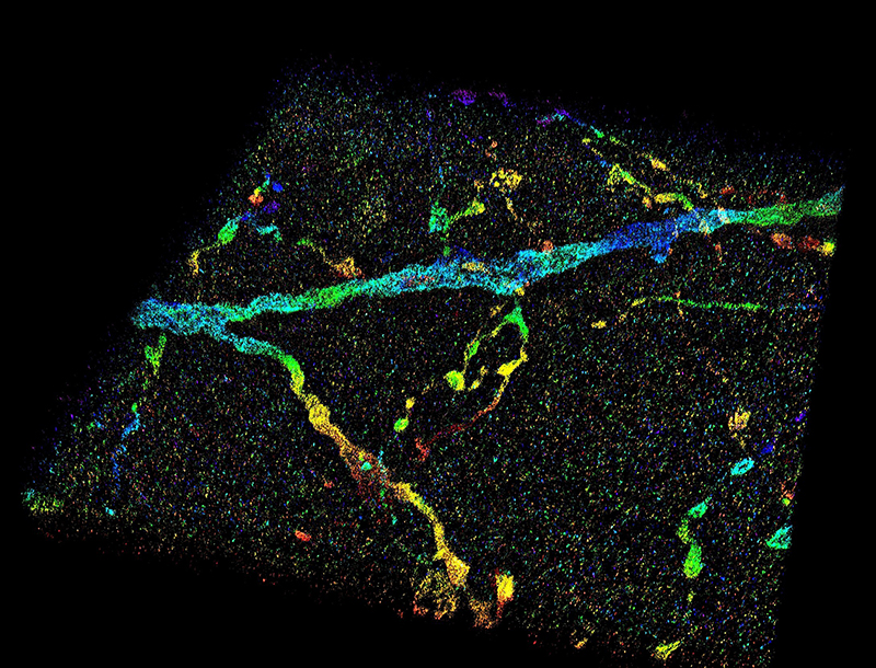 This image shows a 3D super-resolution reconstruction of dendrites in primary visual cortex. Image Credit: Fang Huang/Purdue University.
But resolution, which happens to be the smallest resolvable feature, has been limited by the wave nature of light. This 100-year-old obstacle has limited one’s interpretation of the functions, dynamics, and interactions of cells, especially at the sub-micron to nanometer scale.
This fundamental limit has been resolved by super-resolution fluorescence microscopy, which provides up to 10 times enhancement in terms of resolution, and enables researchers to observe the inner mechanisms of biomolecules and cells at unparalleled spatial resolution.
However, this resolving capability is usually obstructed when visualizing the internal part of whole-cell or whole-tissue specimens, such as the ones that are generally examined during the analyses of the brain or cancer. Molecules within a specimen emit light signals that move via different parts of the tissue or cell structures at varying rates and lead to aberrations, which, in turn, degrade the image.
Scientists from Purdue University have now created a novel technology to resolve this difficulty.
Our technology allows us to measure wavefront distortions induced by the specimen, either a cell or a tissue, directly from the signals generated by single molecules – tiny light sources attached to the cellular structures of interest.
Fang Huang, Assistant Professor of Biomedical Engineering, College of Engineering. Purdue University
Huang continued, “By knowing the distortion induced, we can pinpoint the positions of individual molecules at high precision and accuracy. We obtain thousands to millions of coordinates of individual molecules within a cell or tissue volume and use these coordinates to reveal the nanoscale architectures of specimen constituents.”
The technology developed by the Purdue team was recently published in the Nature Methods journal.
“During three-dimensional super-resolution imaging, we record thousands to millions of emission patterns of single fluorescent molecules,” stated Fan Xu, the study’s co-first author and a postdoctoral associate in Huang’s laboratory.
“These emission patterns can be regarded as random observations at various axial positions sampled from the underlying 3D point-spread function describing the shapes of these emission patterns at different depths, which we aim to retrieve,” Xu added
Xu continued, “Our technology uses two steps: assignment and update, to iteratively retrieve the wavefront distortion and the 3D responses from the recorded single molecule dataset containing emission patterns of molecules at arbitrary locations.”
The new technology devised by the Purdue team might help to identify the locations of biomolecules with an accuracy down to a few nanometers within the whole tissues and cells, and thus overcomes the tissue and cellular designs with excellent fidelity and resolution.
This advancement expands the routine applicability of super-resolution microscopy from selected cellular targets near coverslips to intra- and extra-cellular targets deep inside tissues. This newfound capacity of visualization could allow for better understanding for neurodegenerative diseases such as Alzheimer’s, and many other diseases affecting the brain and various parts inside the body.
Donghan Ma, Study Co-First Author and Postdoctoral Researcher, Purdue University
Donghan Ma works in Huang’s laboratory. The study received major support from the National Institutes of Health.
The research team also includes Gary Landreth, a professor in the School of Medicine at Indiana University; Sarah Calve, an associate professor of biomedical engineering in the College of Engineering at Purdue University (now an associate professor of mechanical engineering at the University of Colorado Boulder); Peng Yin, a Harvard Medical School professor; and Alexander Chubykin, an assistant professor of biological sciences at Purdue University. The entire list of authors is given in the Nature Methods journal.
This technical advancement is startling and will fundamentally change the precision with which we evaluate the pathological features of Alzheimer’s disease. We are able to see smaller and smaller objects and their interactions with each other, which helps reveal structure complexities we have not appreciated before.
Gary Landreth, Professor, School of Medicine, Indiana University
According to Calve, this novel technology represents a major breakthrough in regenerative treatments to help support the repair mechanisms inside the body.
“This development is critical for understanding tissue biology and being able to visualize structural changes,” stated Calve.
According to Chubykin, whose laboratory explores autism and other disorders that impact the brain, the high-resolution imaging technology offers a new technique for interpreting the brain’s impairments.
“This is a tremendous breakthrough in terms of functional and structural analyses,” stated Chubykin. “We can see a much more detailed view of the brain and even mark specific neurons with genetic tools for further study.”
The researchers have patented the technology in association with the Purdue Research Foundation Office of Technology Commercialization.
The office has now shifted to the Convergence Center for Innovation and Collaboration in Discovery Park District, next to Purdue University’s campus. The researchers are looking for partners to commercialize their new imaging technology.
Journal Reference:
Xu, F., et al. (2020) Three-dimensional nanoscopy of whole cells and tissues with in situ point spread function retrieval. Nature Methods. doi.org/10.1038/s41592-020-0816-x.