
Discussion
Independent of the type of sorbent and composition of the eluent, peptide RPLC separation is mostly driven by hydrophobic interactions of amino acid side chains with the nonpolar stationary phase.[15] Therefore, some correlation between peptide retention in any potential pairing of RPLC systems will be observed: analytes containing more hydrophobic residues (Trp, Phe, Leu, Ile, Val, Met and Tyr) will always exhibit higher retention. This results in a characteristic distribution of peptides on orthogonality plots where most of the data lie along a diagonal. Figure 1 IB & IC schematically show the typical dependencies for the RP–RP systems with similar pH, but using a different ion-pairing modifier and for a different pH, respectively. Peptide separations under identical conditions, or using completely different mechanisms compared with RP, will exhibit very poor (Figure 1 IA) or perfect (Figure 1 ID) orthogonality. The importance of separation orthogonality in 2D LC is further illustrated in Figure 1 IIA & IID. One fraction collected in the first dimension will generate very different 'footprints' in the second-dimension RPLC MS run. No distribution of peptides will be observed in a low orthogonality system (Figure 1 IIA), while completely different selectivity will result in perfect utilization of the separation space in the second-dimension RPLC MS (Figure 1 IID), facilitating optimal sample delivery into the mass spectrometer and better identification results.

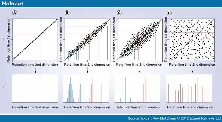
Figure 1.
Separation orthogonality and fraction concatenation in typical 2D reversed-phase–reversed-phase liquid chromatography–mass spectrometry applications. (I) Orthogonality plots for: (A) identical separation conditions in both dimensions; (B) reversed-phase separations at the same pH, but different ion-pairing modifiers [14]; (C) reversed-phase separations at different pH [4,6]; (D) completely orthogonal separations. (II) The distribution of peptides from one fraction in the second dimension: (A) in identical systems with no orthogonality; (B) for a low-orthogonality system using a 4-plex fraction concatenation [14]; (C) in a high-pH/low-pH system with a two-fraction concatenation [6]; (D) in a completely orthogonal system with perfect utilization of separation space in the second dimension.
Finding an optimum first-dimension separation includes a number of additional criteria, such as high separation efficiency and compatibility of eluent systems.[6–8] While SCX separation provides very good orthogonality (similar to Figure 1 ID), the separation efficiency is much lower compared with RPLC. Application of a high salt concentration for peptide elution in SCX mode also creates significant interference with the downstream RPLC MS. RP peptide separation at acidic pH possesses high separation efficiency and compatible eluents, but lacks orthogonality with the standard RPLC using formic/acetic acids as the ion-pairing modifiers, which are suitable for electrospray ionization.
It is already known that a peptide's RP separation selectivity is strongly affected by the eluent pH owing to alteration of the ionization state of ionogenic residues (Arg, Lys, His, Asp and Glu). However, this did not impact mainstream proteomic applications, as most silica-based RP materials show low stability at high pH. The situation has changed following the development of new silica-based stationary phases stable at alkaline pH values: XTerra®/Xbridge™ (Waters) and Luna®/Gemini® (Phenomenex). Gilar et al. showed that a combination of water–acetonitrile gradient at pH 10 and pH 2 exhibits sufficient orthogonality (similar to Figure 1 IC) for effective proteomic fractionations.[4] Similar results were demonstrated for polymer-based monolithic columns by Toll and colleagues.[5] A detailed analysis of high-pH–low-pH orthogonality plots shows that peptides with the largest deviations from the diagonal are mostly basic or acidic.[6,15] Asp and Glu are neutral and moderately hydrophobic in acidic conditions, but at pH 10 they are negatively charged and hydrophilic. The opposite is true for basic residues Arg, Lys and His – they are charged and hydrophilic at pH 2, and neutral/hydrophobic at pH 10. Thus, basic peptides tend to retain more strongly under pH 10 conditions (above the diagonal), while acid peptides are more hydrophilic (below the diagonal in Figure 1 IC).[15]
Figure 1 IIC schematically shows the distribution of a single fraction (labeled in red) collected in the first dimension (high pH) when being separated in the second dimension (low pH). While some distribution of analytes is observed, parts of the chromatogram remain unpopulated. This observation led Dwivedi et al. to develop a concatenation approach: a fraction from the first part of the chromatogram (labeled in green) is added to the later one, and they exhibit very little overlap in the second dimension.[6] This reduced the analysis time by half, with a minimal reduction in protein identification.
Stephanowitz et al. employed a polymeric PLRP-S column with TFA eluent additive in the first dimension.[14] Separation selectivity under these conditions is similar to a C18–formic acid system (schematically shown in Figure 1 IB). Changes are caused by different hydrophobicity of ion-pairing modifiers (TFA vs formic acid) and variation in the nature of the stationary phase (silica-based C18 vs styrene–divinylbenzene copolymer). A single fraction collected in the first dimension will show much tighter distribution in the second dimension (compare the fractions labeled in red in Figure 1 IB & IC). Uniform coverage of the separation space in the second dimension in this case can be corrected by concatenating a larger number of fractions: the authors suggested a 4-plex variant, shown schematically in Figure 1 IIB.
The significant result demonstrated by Stephanowitz et al. is that sufficient separation orthogonality can be achieved using very similar RP separation conditions: differences in separation selectivity might not be as critical to the final result if a proper concatenation procedure is applied.[14] These examples[6,14] show how a better understanding of peptides' RP separation selectivity allows the design of better fractionation procedures to achieve superior output from LC MS.
Expert Rev Proteomics. 2012;9(2):125-128. © 2012 Expert Reviews Ltd.


Comments