An earlier diagnosis to avoid kidney transplants

An analytical technique using high brilliance infrared light produced by the SOLEIL synchrotron has been developed by teams from the CNRS, Paris Sud University, Tenon Hospital in Paris, and the Stoke-on-Trent Cancer Centre (GB) to study the calcification present in the kidneys of patients with renal failure. The results show that it is now possible to identify different types of calcification, some of which are specific to diseases that can be treated. If this information is obtained early, the patients concerned can be treated on time and avoid kidney loss and an eventual kidney transplant.
Chronic renal failure, the end-stage of which is lethal, is increasing dramatically in industrialized countries. While the main cause is type II diabetes, renal crystal deposit diseases can also lead to end-stage renal disease. For the latter, though most forms can be treated medically if diagnosed in time, the only alternative is a kidney transplant. Renal crystal deposit diseases are rare and as a result often misdiagnosed. When a patient suffers from such a disease, without knowing it, and then undergoes a kidney transplant, the new kidney will in turn be affected. Calcifications form and clog the kidney tubules, gradually preventing their function completely, which must then be taken over by dialysis, a very restrictive and major procedure, leading to renal transplantation, if this is possible.
Yet renal crystal deposit diseases are curable by appropriate drug treatments. It is therefore easy to understand the importance of being able to diagnose these in patients, to avoid the necessity of carrying out an operation as serious as a transplant - not to mention the fact that donor tissue is rare.
A unique analysis technique
Such a diagnosis can be carried out on calcifications present in the kidney. Several kinds of calcifications exist: some signal specific crystal deposit diseases, whereas others are the consequences of poor renal function and are not specific to the diseases under investigation. It is therefore necessary to be able to distinguish between them.
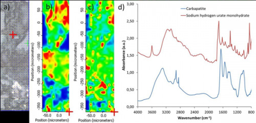
Characterization techniques, based on the differential staining of these calcifications, are already used in hospitals, but these do not differentiate precisely between the kinds of calcifications. In contrast, infrared micro-spectroscopy using synchrotron radiation (SR μFTIR) lifts the uncertainty in a few minutes, and this from samples that may be less than ten microns in size, which is not the case with the conventional FTIR spectroscopy that is available in some laboratories but is not sensitive enough at the scale of the size of the calcifications. Information is obtained as a chemical map of the sample: each pixel of a few square microns on the map provides the precise chemical composition of the calcification.
Groups from CNRS, Paris Sud University, Tenon Hospital in Paris, and the Stoke-on-Trent Cancer Centre (GB) analyzed more than twenty kidney biopsies on the SMIS beamline at SOLEIL synchrotron from patients suffering from various kidney diseases. Due to the intensity of infrared radiation and the microscopic size of the light spot analyzing the samples, the researchers managed to identify several types of crystals, the composition of which was observed in some cases for the first time. Another discovery: a single biopsy can contain two or three different crystalline phases. This is the first scientific evidence of the diversity and heterogeneity of calcifications found in tissues.
These results show that the SRμFTIR technique is an exceptional and unique tool for analyzing calcifications associated with kidney alteration. Hopefully they will bring great improvements in the early diagnosis and treatment of some kidney diseases, so far treated by often repeat kidney transplants, because of the destruction of one or more successive grafts by the unidentified crystal deposits.
More information: Shedding light on the chemical diversity of ectopic calcifications in kidney tissues: Diagnostic and research aspects,A. Dessombz et al., PLoS ONE (2011).


