Scientists at Texas Heart Institute (THI) and Rice University have used biocompatible fibres made of carbon nanotubes (CNTs) as electrical bridges to restore conductivity to damaged hearts. By sewing the fibres directly into damaged cardiac tissue in animal models, they could deliver the electrical signals needed to keep the animals’ hearts beating (Circ. Arrhythm Electrophysiol. 10.1161/CIRCEP.119.007256).
“Instead of shocking and defibrillating, we are actually correcting diseased conduction of the largest major pumping chamber of the heart by creating a bridge to bypass and conduct over a scarred area of a damaged heart,” explains THI’s Mehdi Razavi, who co-led the study with Matteo Pasquali from Rice.
“Today there is no technology that treats the underlying cause of the number one cause of sudden death – ventricular arrhythmias,” Razavi adds. “These arrhythmias are caused by the disorganized firing of impulses from the heart’s lower chambers and are challenging to treat in patients after a heart attack or with scarred heart tissue due to such other conditions as congestive heart failure or dilated cardiomyopathy.”
And while many effective antiarrhythmic drugs are available, they are often contraindicated in patients after a heart attack. What’s really needed therapeutically, is a way to increase conduction.
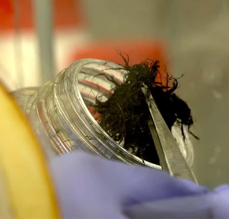
To achieve this, the researchers employed CNT fibres invented by Pasquali’s lab. The fibres combine the mechanical properties of suture materials with the conductive properties of metals. The team demonstrated that the polymer-coated fibres, with their ends stripped to create electrodes, could restore function in sheep and rodents with damaged hearts, whether the initial conduction was slowed, severed or blocked.
The researchers used radiofrequency ablation to create epicardial conduction delay in sheep and then applied CNT fibres. They found that sewing the conductive fibres across the ablation-induced scar significantly improved conduction, to near baseline values.
In an acute electrophysiology study on rodents, the fibres restored myocardial conduction across scar in sinus atrial rhythm without controlled external pacing. In a chronic study, the fibres maintained conduction for one month after atrioventricular nodal ablation, but required atrial pacing. The researchers note that no gross or histopathologic evidence of toxicity was observed.
“Our experiments provided the first scientific support for using a synthetic material-based treatment rather than a drug to treat the leading cause of sudden death in the US and many developing countries around the world,” says Razavi.
Many questions remain, however, before the procedure can move toward human testing. The researchers must establish a way to sew the fibres in place using a minimally invasive catheter, and ensure that the fibres are strong and flexible enough to serve a constantly beating heart over the long term. The team also needs to determine how long and wide the fibres should be, precisely how much electricity they need to carry and how they would perform in the growing hearts of young patients.
“Flexibility is important because the heart is continuously pulsating and moving, so anything that’s attached to the heart’s surface is going to be deformed and flexed,” says Pasquali. “Good interfacial contact is also critical to pick up and deliver the electrical signal,” he said. “In the past, multiple materials had to be combined to attain both electrical conductivity and effective contacts. These fibres have both properties built in by design, which greatly simplifies device construction and lowers risks of long-term failure due to delamination of multiple layers or coatings.”